Copyright
©The Author(s) 2020.
World J Orthop. Nov 18, 2020; 11(11): 475-482
Published online Nov 18, 2020. doi: 10.5312/wjo.v11.i11.475
Published online Nov 18, 2020. doi: 10.5312/wjo.v11.i11.475
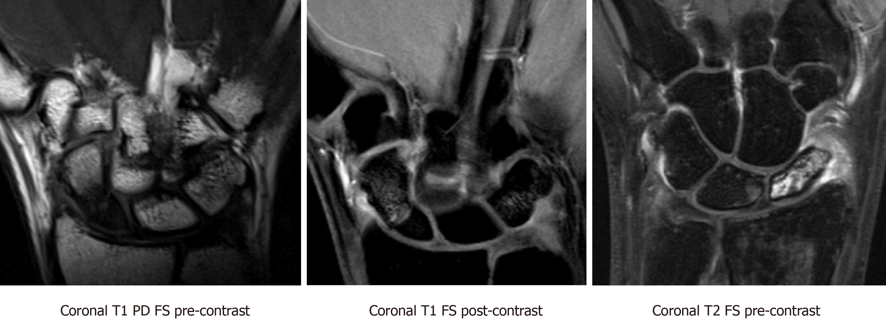
Figure 7 Coronal T1-weighted proton density fat saturation pre-contrast, Coronal T1-weighted fat saturation post-contrast, and coronal T2-weighted fat saturation pre-contrast magnetic resonance imaging sequences demonstrating nondisplaced ununited fracture of the proximal pole of the scaphoid.
There is T1 hypointense and heterogeneous T2 signal within the proximal pole fracture fragment. The proximal pole fracture fragment demonstrates no significant contrast enhancement on the postcontrast images, compatible with avascular necrosis. PD: Proton density; FS: Fat saturation.
- Citation: Cheema HS, Cheema AN. Radiographic evaluation of vascularity in scaphoid nonunions: A review. World J Orthop 2020; 11(11): 475-482
- URL: https://www.wjgnet.com/2218-5836/full/v11/i11/475.htm
- DOI: https://dx.doi.org/10.5312/wjo.v11.i11.475


