Copyright
©The Author(s) 2020.
World J Orthop. Nov 18, 2020; 11(11): 475-482
Published online Nov 18, 2020. doi: 10.5312/wjo.v11.i11.475
Published online Nov 18, 2020. doi: 10.5312/wjo.v11.i11.475
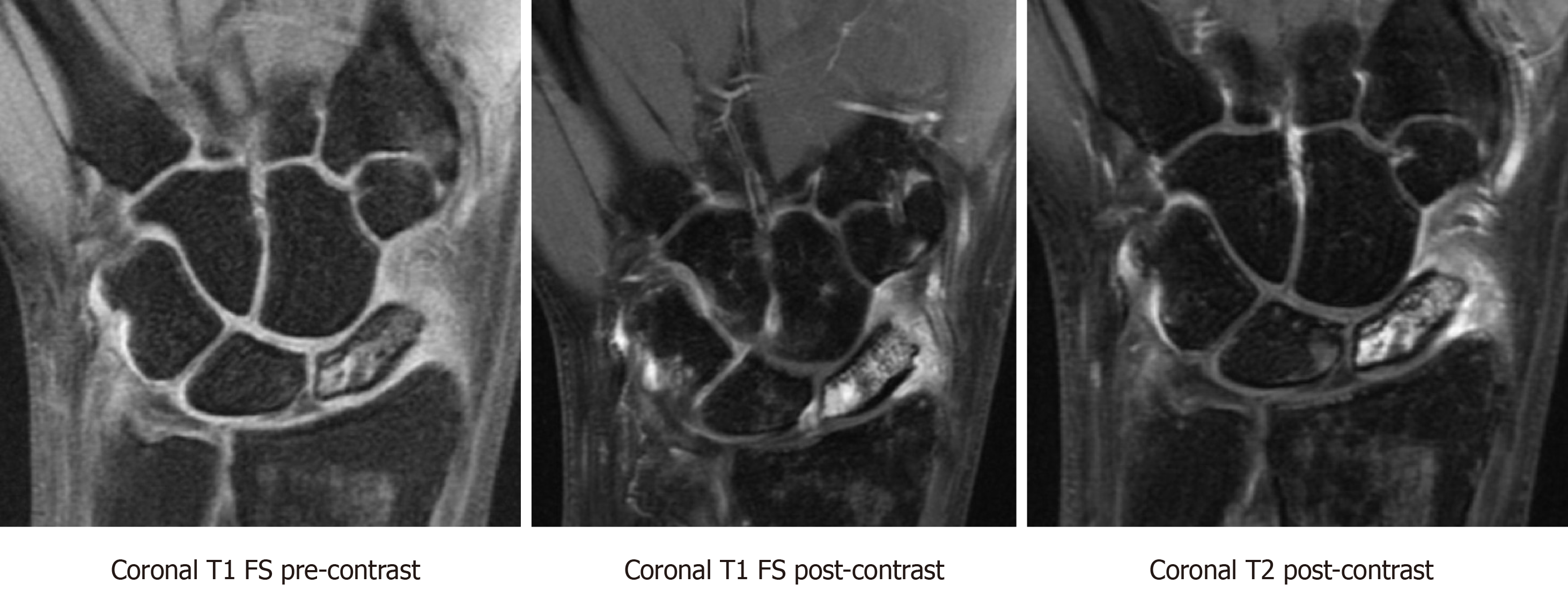
Figure 6 T1-weighted fat saturated pre-contrast, T1-weighted fat saturated post-contrast, and T2-weighted post-contrast sequences showing preserved vascularity in a proximal pole scaphoid fracture.
There is a hypointense fracture line and diffuse marrow edema throughout the scaphoid. There is also hypointense T1 signal and hyperintense T2 signal with corresponding hyperenhancement on post-contrast imaging at the proximal pole, most intense in the proximal pole in keeping with preserved vascularity. FS: Fat saturated.
- Citation: Cheema HS, Cheema AN. Radiographic evaluation of vascularity in scaphoid nonunions: A review. World J Orthop 2020; 11(11): 475-482
- URL: https://www.wjgnet.com/2218-5836/full/v11/i11/475.htm
- DOI: https://dx.doi.org/10.5312/wjo.v11.i11.475


