Copyright
©The Author(s) 2020.
World J Orthop. Nov 18, 2020; 11(11): 475-482
Published online Nov 18, 2020. doi: 10.5312/wjo.v11.i11.475
Published online Nov 18, 2020. doi: 10.5312/wjo.v11.i11.475
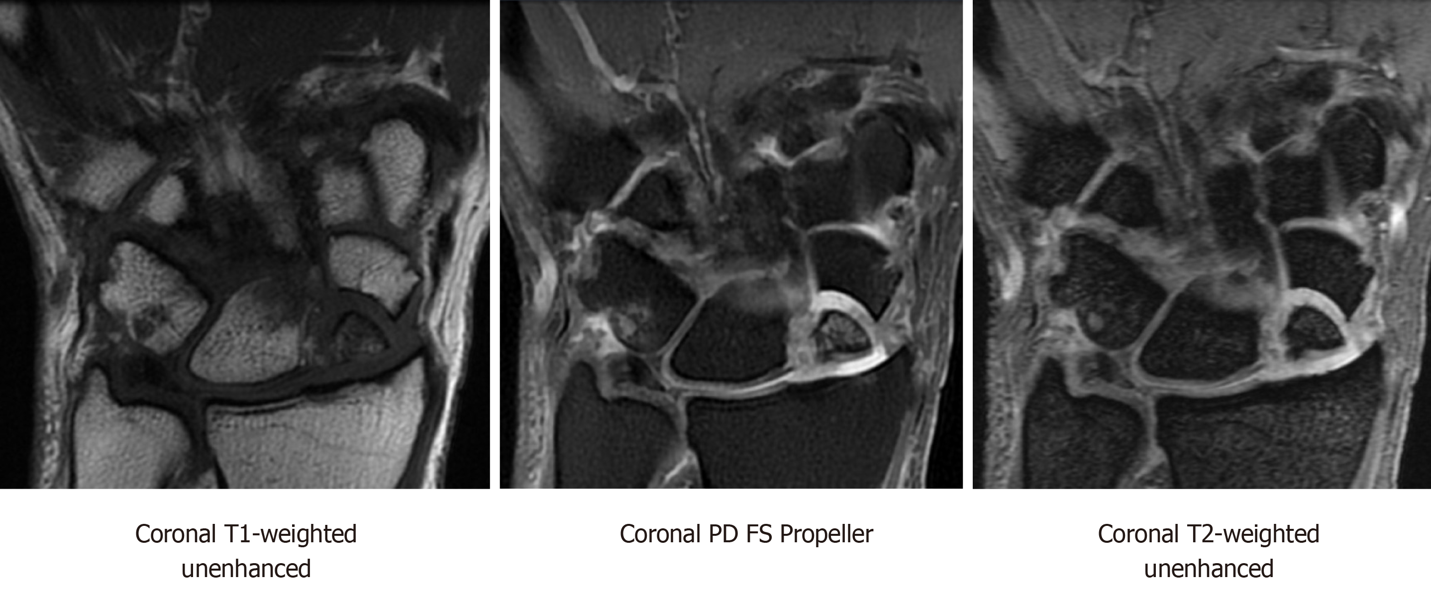
Figure 2 Coronal magnetic resonance imaging T1-weighted unenhanced, coronal proton density fat-saturation propeller, and coronal T2-weighted sequences showing scaphoid nonunion advanced collapse with chronic ununited comminuted scaphoid fracture involving the proximal pole and mid waist.
There are several displaced and necrotic proximal pole fragments, and severe chondrosis at the radioscaphoid articulation. PD: Proton density; FS: Fat-saturation.
- Citation: Cheema HS, Cheema AN. Radiographic evaluation of vascularity in scaphoid nonunions: A review. World J Orthop 2020; 11(11): 475-482
- URL: https://www.wjgnet.com/2218-5836/full/v11/i11/475.htm
- DOI: https://dx.doi.org/10.5312/wjo.v11.i11.475


