Copyright
©The Author(s) 2019.
World J Orthop. Jul 18, 2019; 10(7): 292-298
Published online Jul 18, 2019. doi: 10.5312/wjo.v10.i7.292
Published online Jul 18, 2019. doi: 10.5312/wjo.v10.i7.292
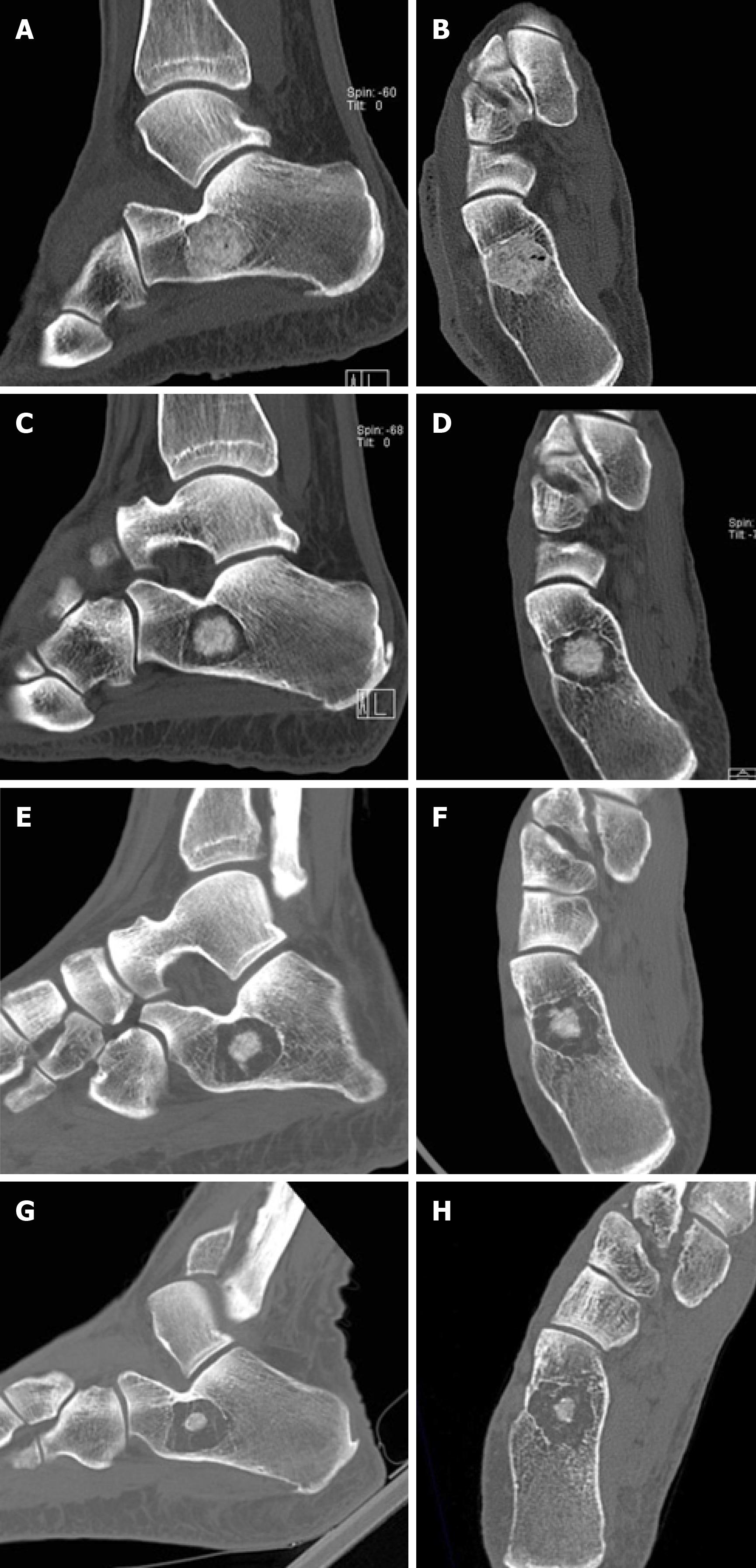
Figure 2 Serial computed tomography evaluation of os calcis lesion.
A and B: Computed tomography images immediately postoperatively. Complete filling of the cavity has been obtained with bone graft. C and D: Six months postoperatively, circumferential graft absorption is present. E and F: Fifteen months postoperatively, further graft absorption is noted. G and H: At five years, the greatest part of the bone graft has been resorbed. A mesh of thin osseous septa is seen around the remaining bone graft. The lesion has not expanded since the first postoperative image.
- Citation: Balbouzis T, Alexopoulos T, Grigoris P. Os calcis lipoma: To graft or not to graft? - A case report and literature review. World J Orthop 2019; 10(7): 292-298
- URL: https://www.wjgnet.com/2218-5836/full/v10/i7/292.htm
- DOI: https://dx.doi.org/10.5312/wjo.v10.i7.292


