Copyright
©The Author(s) 2019.
World J Orthop. Jan 18, 2019; 10(1): 45-53
Published online Jan 18, 2019. doi: 10.5312/wjo.v10.i1.45
Published online Jan 18, 2019. doi: 10.5312/wjo.v10.i1.45
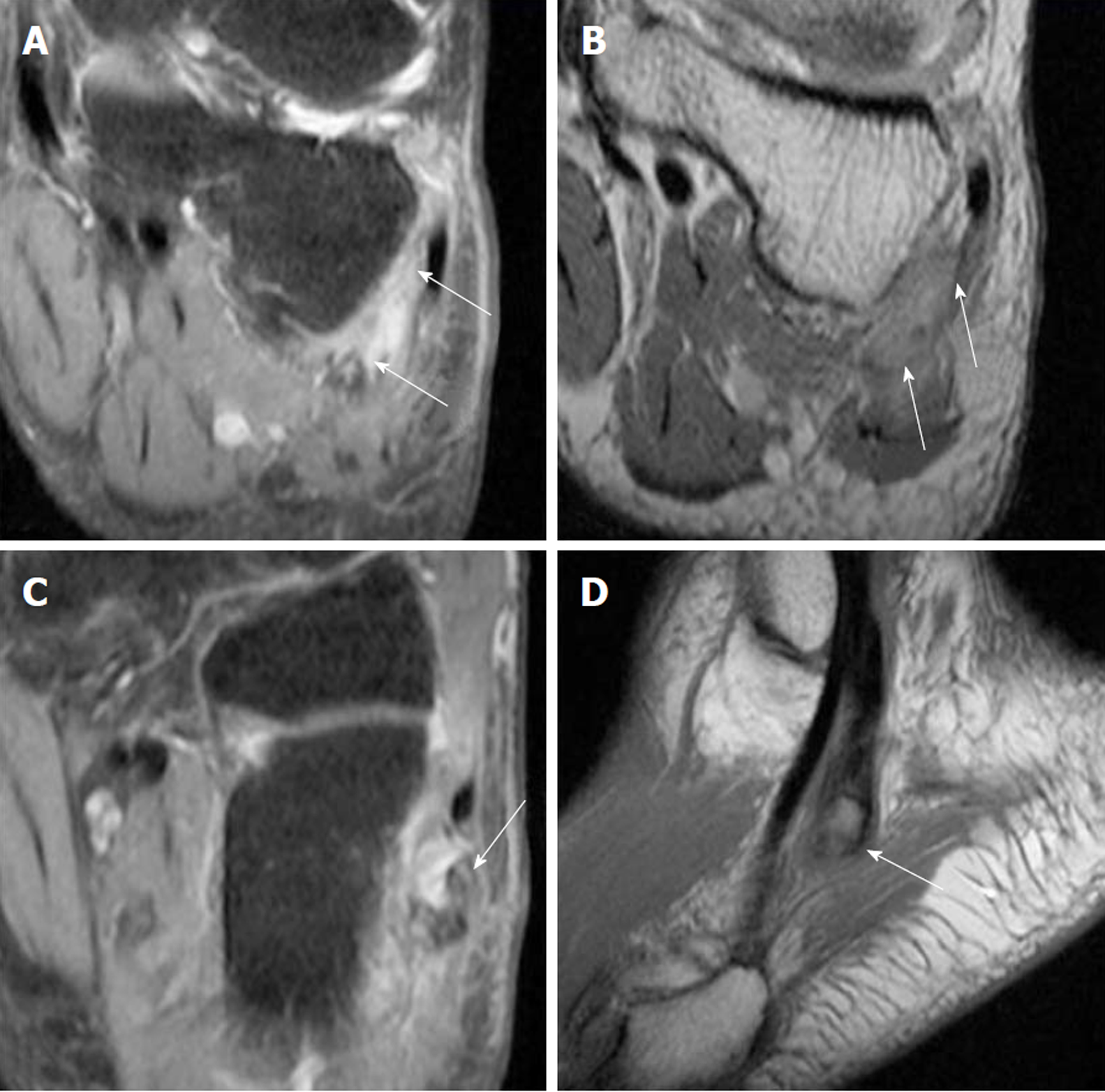
Figure 3 Magnetic resonance imaging of left foot.
A: (Coronal) Peroneal tenosynovitis; B: (Coronal) Both peroneal tendons run within a flat peroneal groove with intact superior peroneal retinaculum. No fixed lateral subluxation of the peroneal tendons; C, D: (Sagittal view). Five millimeter os peroneum within the peroneus longus tendon (PLT), with full-thickness rupture of the PLT. The tear measures 2 cm in length, with proximal migration of the tendon at the level of the cuboid bone.
- Citation: Koh D, Liow L, Cheah J, Koo K. Peroneus longus tendon rupture: A case report. World J Orthop 2019; 10(1): 45-53
- URL: https://www.wjgnet.com/2218-5836/full/v10/i1/45.htm
- DOI: https://dx.doi.org/10.5312/wjo.v10.i1.45


