Copyright
©The Author(s) 2018.
World J Clin Oncol. Dec 20, 2018; 9(8): 200-207
Published online Dec 20, 2018. doi: 10.5306/wjco.v9.i8.200
Published online Dec 20, 2018. doi: 10.5306/wjco.v9.i8.200
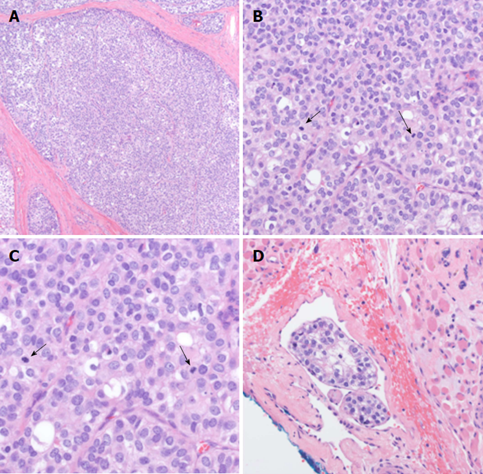
Figure 2 Hematoxylin and eosin stain of epithelial-myoepithelial carcinoma specimen.
A: Epithelial-myoepithelial carcinoma (EMC), 10 × magnification; B: EMC, 40 × magnification; C: EMC, 60 × magnification; D: Lymphovasuclar invasion, 40 × magnification. Arrows indicate mitotic figures.
- Citation: Khattab MH, Sherry AD, Ahlers CG, Kirschner AN. Radiation-associated epithelial-myoepithelial carcinoma among five secondary malignancies: A case report and review of literature. World J Clin Oncol 2018; 9(8): 200-207
- URL: https://www.wjgnet.com/2218-4333/full/v9/i8/200.htm
- DOI: https://dx.doi.org/10.5306/wjco.v9.i8.200


