Copyright
©The Author(s) 2018.
World J Clin Oncol. Nov 10, 2018; 9(7): 162-166
Published online Nov 10, 2018. doi: 10.5306/wjco.v9.i7.162
Published online Nov 10, 2018. doi: 10.5306/wjco.v9.i7.162
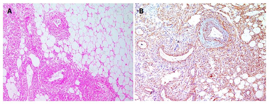
Figure 4 Final histopathology of surgical specimen.
A: Components of all the three tissues, i.e., mature adipocytes, spindle shaped smooth muscle cells and blood vessel (10 ×); B: Immunohistochemical staining for smooth muscle actin shows strong positivity in final histology specimen.
- Citation: Sharma G, Jain A, Sharma P, Sharma S, Rathi V, Garg PK. Giant exophytic renal angiomyolipoma masquerading as a retroperitoneal liposarcoma: A case report and review of literature. World J Clin Oncol 2018; 9(7): 162-166
- URL: https://www.wjgnet.com/2218-4333/full/v9/i7/162.htm
- DOI: https://dx.doi.org/10.5306/wjco.v9.i7.162


