Copyright
©The Author(s) 2018.
World J Clin Oncol. Nov 10, 2018; 9(7): 162-166
Published online Nov 10, 2018. doi: 10.5306/wjco.v9.i7.162
Published online Nov 10, 2018. doi: 10.5306/wjco.v9.i7.162
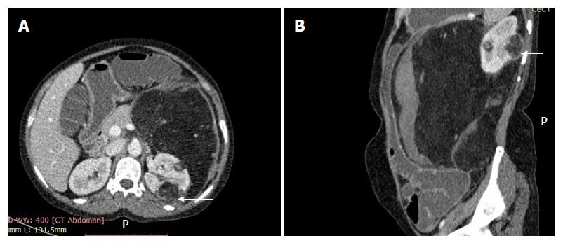
Figure 1 Axial section of contrast enhancement computed tomography abdomen.
A: It shows two fairly well defined, predominantly fat density rounded lesions, in the left kidney. The peripheral capsule (arrow) of the smaller lesion is ill defined and continuous with a very large perirenal angiomyolipoma showing prominent vessels (+); B: Sagittal reconstruction shows a peripheral hematoma (arrow) in the perirenal lipomatous mass.
- Citation: Sharma G, Jain A, Sharma P, Sharma S, Rathi V, Garg PK. Giant exophytic renal angiomyolipoma masquerading as a retroperitoneal liposarcoma: A case report and review of literature. World J Clin Oncol 2018; 9(7): 162-166
- URL: https://www.wjgnet.com/2218-4333/full/v9/i7/162.htm
- DOI: https://dx.doi.org/10.5306/wjco.v9.i7.162


