Copyright
©The Author(s) 2018.
World J Clin Oncol. Feb 10, 2018; 9(1): 20-25
Published online Feb 10, 2018. doi: 10.5306/wjco.v9.i1.20
Published online Feb 10, 2018. doi: 10.5306/wjco.v9.i1.20
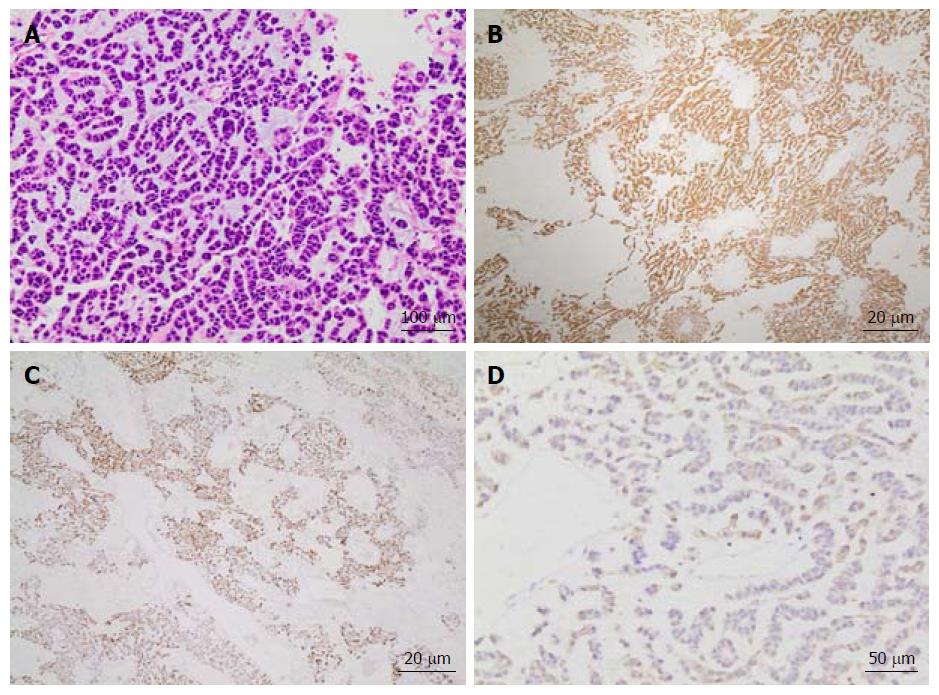
Figure 1 Tissue histopathology of a hepatic lesion confirms metastatic adrenocortical carcinoma.
Hematoxylin and eosin (HE) staining (A) at 200 × magnification of tissue from the hepatic mass shows diffuse infiltrating cords of cells with hyperchromasia, eosinophilic cytoplasm, and a mild degree of nuclear pleomorphism in a background of myxoid stroma. Additional positive immunostaining with inhibin (B) and melan-A (C) at 40 × magnification as well as CKAE1/3 (D) at 100 × magnification confirms the diagnosis as adrenocortical carcinoma.
- Citation: Makary MS, Krishner LS, Wuthrick EJ, Bloomston MP, Dowell JD. Yttrium-90 microsphere selective internal radiation therapy for liver metastases following systemic chemotherapy and surgical resection for metastatic adrenocortical carcinoma. World J Clin Oncol 2018; 9(1): 20-25
- URL: https://www.wjgnet.com/2218-4333/full/v9/i1/20.htm
- DOI: https://dx.doi.org/10.5306/wjco.v9.i1.20


