Copyright
©The Author(s) 2017.
World J Clin Oncol. Aug 10, 2017; 8(4): 366-370
Published online Aug 10, 2017. doi: 10.5306/wjco.v8.i4.366
Published online Aug 10, 2017. doi: 10.5306/wjco.v8.i4.366
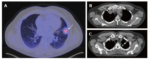
Figure 3 Positron emission tomography–computed tomography, three months later.
A: Subpleural hypermetabolic nodular lesion in left upper lobe (SUV max 3.8) measuring 27 mm × 30 mm, suggestive of tumor activity; B and C: Computed tomography, a 38-mm nodule in right upper lobe and another 20-mm nodule in left upper lobe, compatible with metastasis.
- Citation: García-Cabezas S, Centeno-Haro M, Espejo-Pérez S, Carmona-Asenjo E, Moreno-Vega AL, Ortega-Salas R, Palacios-Eito A. Intimal sarcoma of the pulmonary artery with multiple lung metastases: Long-term survival case. World J Clin Oncol 2017; 8(4): 366-370
- URL: https://www.wjgnet.com/2218-4333/full/v8/i4/366.htm
- DOI: https://dx.doi.org/10.5306/wjco.v8.i4.366


