Copyright
©The Author(s) 2017.
World J Clin Oncol. Aug 10, 2017; 8(4): 366-370
Published online Aug 10, 2017. doi: 10.5306/wjco.v8.i4.366
Published online Aug 10, 2017. doi: 10.5306/wjco.v8.i4.366
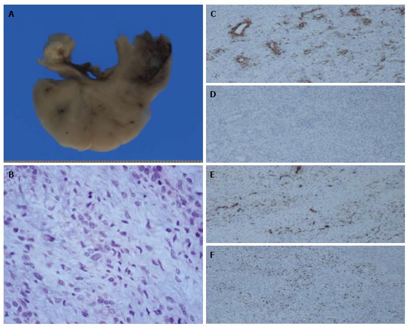
Figure 2 The patient was operated on for a suspected primary tumor of the pulmonary artery, resulting in with complete resection of the mass, whose pathological result was an intermediate-grade malignant tumor suggestive of Pulmonary artery intimal sarcoma.
A: Macro: View of the pulmonary artery transversal section with infiltrating sarcoma on the lumen; B: High power of the tumor. Note the variable atypia 200 ×. Immunohistochemical stainings 100 ×; C: Smooth muscle actin: Focal tumor cell reaction 100 ×; D: Desmine: Negative 100 ×; E: CD31: Reaction of the endothelium surrounded by negative tumor cells 100 ×; F: Ki-67: Variable proliferation index in tumor cells 100 ×.
- Citation: García-Cabezas S, Centeno-Haro M, Espejo-Pérez S, Carmona-Asenjo E, Moreno-Vega AL, Ortega-Salas R, Palacios-Eito A. Intimal sarcoma of the pulmonary artery with multiple lung metastases: Long-term survival case. World J Clin Oncol 2017; 8(4): 366-370
- URL: https://www.wjgnet.com/2218-4333/full/v8/i4/366.htm
- DOI: https://dx.doi.org/10.5306/wjco.v8.i4.366


