Copyright
©The Author(s) 2017.
World J Clin Oncol. Aug 10, 2017; 8(4): 305-319
Published online Aug 10, 2017. doi: 10.5306/wjco.v8.i4.305
Published online Aug 10, 2017. doi: 10.5306/wjco.v8.i4.305
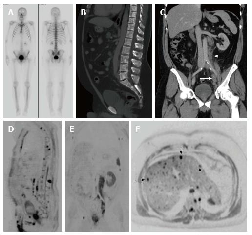
Figure 8 A 51-year-old patient with a history of Gleason 4 + 5 prostate carcinoma treated with hormone therapy.
A: Re-staging bone scintigraphy was negative for bone metastases; B and C: Sagittal (B) and coronal (C) images of abdominal CT scan show a diffuse axial bone altered density that could not rule out bone metastases, along with multiple retroperitoneal and pelvic enlarged nodes suggesting malignant adenopathies (white arrows in C); D and E: Sagittal (D) and coronal (E) whole-body MRI images clearly show multiple bone metastases and adenopathies, along with hepatic nodules (black arrows in F, axial abdominal MRI) suspicious for metastatic disease. MRI: Magnetic resonance imaging.
- Citation: Couñago F, Sancho G, Catalá V, Hernández D, Recio M, Montemuiño S, Hernández JA, Maldonado A, del Cerro E. Magnetic resonance imaging for prostate cancer before radical and salvage radiotherapy: What radiation oncologists need to know. World J Clin Oncol 2017; 8(4): 305-319
- URL: https://www.wjgnet.com/2218-4333/full/v8/i4/305.htm
- DOI: https://dx.doi.org/10.5306/wjco.v8.i4.305


