Copyright
©The Author(s) 2017.
World J Clin Oncol. Aug 10, 2017; 8(4): 305-319
Published online Aug 10, 2017. doi: 10.5306/wjco.v8.i4.305
Published online Aug 10, 2017. doi: 10.5306/wjco.v8.i4.305
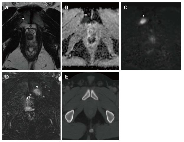
Figure 7 A 54-year-old patient with recently diagnosed prostate carcinoma.
A: T2-weighted axial image of the pelvis shows an ill-defined hyperintense area in the right pubis (arrow); B and C: ADC map (B) and DWI images (C) show restriction of diffusion in the same area (arrow); D: DCE image presents enhancement of the lesion after IV gadolinium (arrow). MRI findings were suspicious for pelvic bone metastasis, with no evidence of such lesion on staging abdomino-pelvic CT scan performed a week earlier (E) and in a previous bone scintigraphy (not shown). Bone metastasis was confirmed on clinical evolution. MRI: Magnetic resonance imaging; DCE: Dynamic contrast-enhanced; CT: Computed tomography.
- Citation: Couñago F, Sancho G, Catalá V, Hernández D, Recio M, Montemuiño S, Hernández JA, Maldonado A, del Cerro E. Magnetic resonance imaging for prostate cancer before radical and salvage radiotherapy: What radiation oncologists need to know. World J Clin Oncol 2017; 8(4): 305-319
- URL: https://www.wjgnet.com/2218-4333/full/v8/i4/305.htm
- DOI: https://dx.doi.org/10.5306/wjco.v8.i4.305


