Copyright
©The Author(s) 2017.
World J Clin Oncol. Aug 10, 2017; 8(4): 305-319
Published online Aug 10, 2017. doi: 10.5306/wjco.v8.i4.305
Published online Aug 10, 2017. doi: 10.5306/wjco.v8.i4.305
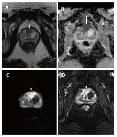
Figure 3 A 71-year-old patient.
A: T2-weighted axial image at the level of the midgland of the prostate shows a hypointense nodular lesion at the transitional zone/anterior fibromuscular stroma, with a diameter of 26 mm (arrow); B and C: ADC map (B) and DWI image (C) show a marked hypo- and hyperintensity, respectively, in relation to restriction of diffusion (white arrows); D: DCE image with significant enhancement of the lesion (arrow). The characteristics of the nodule are compatible with a PIRADS 5 lesion and the marked restriction of the diffusion suggests a high-grade clinically significant prostate carcinoma, confirmed by the results of a MRI-guided transrectal ultrasound prostate biopsy (Gleason 4 + 4).
- Citation: Couñago F, Sancho G, Catalá V, Hernández D, Recio M, Montemuiño S, Hernández JA, Maldonado A, del Cerro E. Magnetic resonance imaging for prostate cancer before radical and salvage radiotherapy: What radiation oncologists need to know. World J Clin Oncol 2017; 8(4): 305-319
- URL: https://www.wjgnet.com/2218-4333/full/v8/i4/305.htm
- DOI: https://dx.doi.org/10.5306/wjco.v8.i4.305


