Copyright
©The Author(s) 2017.
World J Clin Oncol. Jun 10, 2017; 8(3): 293-299
Published online Jun 10, 2017. doi: 10.5306/wjco.v8.i3.293
Published online Jun 10, 2017. doi: 10.5306/wjco.v8.i3.293
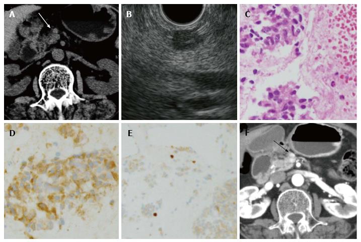
Figure 3 Pancreatic neuroendocrine tumor case followed up without surgery.
A: Abdominal CT. A tumor was recognized in the pancreatic body. The diameter of the lesion was 8 mm; B: Endoscopic ultrasonography. The tumor was recognized as a low echoic lesion. A 22G needle was inserted into the tumor; C: Hematoxylin and eosin stain (× 400). Spindle-shaped tumor cells with ellipse nuclei formed funicular lines; D: Chromogranin A staining (× 400). Tumor cells were chromogranin A-positive; E: The Ki-67 index was < 1.0% (× 200), with tumor grade G1; F: Abdominal CT. The tumor did not grow after 54 mo. PNET: Pancreatic neuroendocrine tumor; CT: Computed tomography.
- Citation: Sugimoto M, Takagi T, Suzuki R, Konno N, Asama H, Watanabe K, Nakamura J, Kikuchi H, Waragai Y, Takasumi M, Kawana S, Hashimoto Y, Hikichi T, Ohira H. Pancreatic neuroendocrine tumor Grade 1 patients followed up without surgery: Case series. World J Clin Oncol 2017; 8(3): 293-299
- URL: https://www.wjgnet.com/2218-4333/full/v8/i3/293.htm
- DOI: https://dx.doi.org/10.5306/wjco.v8.i3.293


