Copyright
©The Author(s) 2017.
World J Clin Oncol. Jun 10, 2017; 8(3): 293-299
Published online Jun 10, 2017. doi: 10.5306/wjco.v8.i3.293
Published online Jun 10, 2017. doi: 10.5306/wjco.v8.i3.293
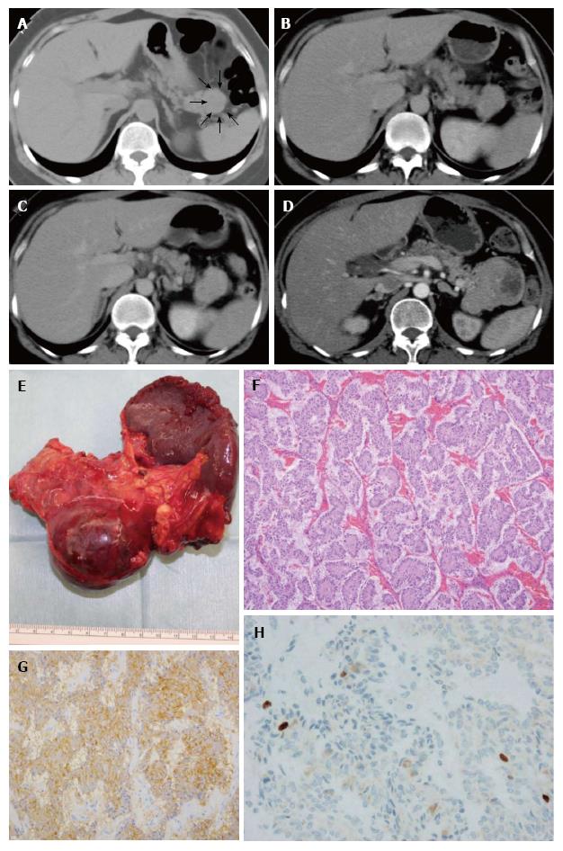
Figure 2 The patient who exhibited growth of the pancreatic neuroendocrine tumor.
A: Abdominal CT. Initial CT indicated a PNET. The lesion was identified in the pancreatic tail. The diameter of the PNET was 34 mm (arrow); B: The lesion grew slightly after 11 mo; C: The lesion grew further after 29 mo; D: The diameter of the tumor became larger than 70 mm after 79 mo; E: The patient underwent distal pancreatectomy after 80 mo; F: Hematoxylin and eosin stain (× 100). Tumor cells formed ribbon-like lines; G: Chromogranin A staining (× 200). Tumor cells were chromogranin A positive; H: The Ki-67 index was 0.9%, with tumor grade G1 (× 200). PNET: Pancreatic neuroendocrine tumor; CT: Computed tomography.
- Citation: Sugimoto M, Takagi T, Suzuki R, Konno N, Asama H, Watanabe K, Nakamura J, Kikuchi H, Waragai Y, Takasumi M, Kawana S, Hashimoto Y, Hikichi T, Ohira H. Pancreatic neuroendocrine tumor Grade 1 patients followed up without surgery: Case series. World J Clin Oncol 2017; 8(3): 293-299
- URL: https://www.wjgnet.com/2218-4333/full/v8/i3/293.htm
- DOI: https://dx.doi.org/10.5306/wjco.v8.i3.293


