Copyright
©The Author(s) 2017.
World J Clin Oncol. Jun 10, 2017; 8(3): 214-229
Published online Jun 10, 2017. doi: 10.5306/wjco.v8.i3.214
Published online Jun 10, 2017. doi: 10.5306/wjco.v8.i3.214
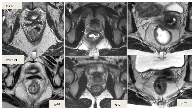
Figure 17 Tumor restaging after neoadjuvant chemoradiotherapy.
On T2-weighted MR images in different patients showing baseline and post-CRT images on upper and lower series, respectively. A: In ypT0 rectal tumor, posttreatment axial image shows a normal, two-layered rectal wall (arrow), corresponding to complete response; B: In ypT3 rectal tumor, posttreatment axial image shows normal, two-layered rectal wall (arrow). This is an example for false-negative MR assessment of complete tumor regression; C: In ypT0 rectal tumor, posttreatment axial image shows thick, fibrotic low signal intensity scar (arrow) in pretreatment T3 tumor area. CRT: Chemoradiotherapy.
- Citation: Engin G, Sharifov R. Magnetic resonance imaging for diagnosis and neoadjuvant treatment evaluation in locally advanced rectal cancer: A pictorial review. World J Clin Oncol 2017; 8(3): 214-229
- URL: https://www.wjgnet.com/2218-4333/full/v8/i3/214.htm
- DOI: https://dx.doi.org/10.5306/wjco.v8.i3.214


