Copyright
©The Author(s) 2017.
World J Clin Oncol. Jun 10, 2017; 8(3): 214-229
Published online Jun 10, 2017. doi: 10.5306/wjco.v8.i3.214
Published online Jun 10, 2017. doi: 10.5306/wjco.v8.i3.214
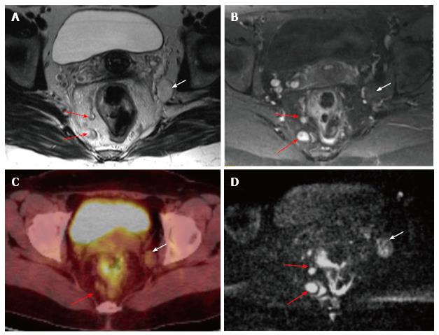
Figure 16 Mesorectal and extramesorectal lymph node involvement in rectal cancer.
A: T2-weighted; B: T1-weighted contrast-enhanced axial MR images; C: 18F-FDG PET-CT; D: DWI showing suspicious lymph nodes in mesorectal (red arrows) and extramesorectal areas (white areas). On DWI, extramesorectal lymph node is more remarkable than T2W and contrast-enhanced T1W sequences. DWI: Diffusion-weighted imaging; 18F-FDG PET-CT: 18F-fluorodeoxyglucosepositron emission tomography-computedtomography.
- Citation: Engin G, Sharifov R. Magnetic resonance imaging for diagnosis and neoadjuvant treatment evaluation in locally advanced rectal cancer: A pictorial review. World J Clin Oncol 2017; 8(3): 214-229
- URL: https://www.wjgnet.com/2218-4333/full/v8/i3/214.htm
- DOI: https://dx.doi.org/10.5306/wjco.v8.i3.214


