Copyright
©The Author(s) 2017.
World J Clin Oncol. Jun 10, 2017; 8(3): 214-229
Published online Jun 10, 2017. doi: 10.5306/wjco.v8.i3.214
Published online Jun 10, 2017. doi: 10.5306/wjco.v8.i3.214
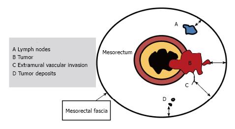
Figure 11 Schematic representation of positive resection margin.
For T3 tumors, the shortest distance between the most penetrating parts of the tumor and the MRF is measured (double-headed arrows). A tumor mesorectal fascia distance of more than 1 mm is a reliable predictor for negative margins. In the presence of satellite nodules such as tumor deposits, lymph nodes or EMVI the shortest distance between the nodules and the MRF should also be reported (Adapted from ref. [27]: Nougaret S, Reinhold C, Mikhael HW, Rouanet P, Bibeau F, Brown G. The use of MR imaging in treatment planning for patients with rectal carcinoma: have you checked the “DISTANCE”? Radiology 2013; 268: 330-344). EMVI: Extramural vascular invasion; MRF: Mesorectal fascia.
- Citation: Engin G, Sharifov R. Magnetic resonance imaging for diagnosis and neoadjuvant treatment evaluation in locally advanced rectal cancer: A pictorial review. World J Clin Oncol 2017; 8(3): 214-229
- URL: https://www.wjgnet.com/2218-4333/full/v8/i3/214.htm
- DOI: https://dx.doi.org/10.5306/wjco.v8.i3.214


