Copyright
©The Author(s) 2017.
World J Clin Oncol. Jun 10, 2017; 8(3): 214-229
Published online Jun 10, 2017. doi: 10.5306/wjco.v8.i3.214
Published online Jun 10, 2017. doi: 10.5306/wjco.v8.i3.214
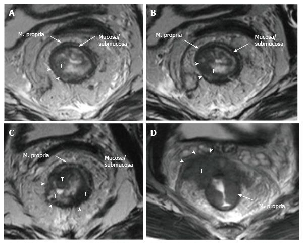
Figure 9 Rectal cancer T staging on magnetic resonance imaging.
T2-weighted axial images showing rectal carcinomas with different T stages. A: T1 tumor is confined to the submucosa, has not entered the muscularis propria (arrowheads); B: T2 tumor extends into, but not beyond, the muscularis propria (arrowheads); C: T3 tumor extends beyond the muscularis propria and strands into mesorectal fat (arrowheads); D: T4a tumor invades the visceral peritoneum (arrowheads). T: Tumor.
- Citation: Engin G, Sharifov R. Magnetic resonance imaging for diagnosis and neoadjuvant treatment evaluation in locally advanced rectal cancer: A pictorial review. World J Clin Oncol 2017; 8(3): 214-229
- URL: https://www.wjgnet.com/2218-4333/full/v8/i3/214.htm
- DOI: https://dx.doi.org/10.5306/wjco.v8.i3.214


