Copyright
©The Author(s) 2017.
World J Clin Oncol. Jun 10, 2017; 8(3): 214-229
Published online Jun 10, 2017. doi: 10.5306/wjco.v8.i3.214
Published online Jun 10, 2017. doi: 10.5306/wjco.v8.i3.214
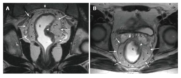
Figure 6 Magnetic resonance imaging anatomy of mesorectum and mesorectal fascia.
On T2-weighted (A) axial and (B) coronal plane magnetic resonance images, mesorectal fascia (arrows) is seen as a thin, low-signal intensity layer enveloping the mesorectal fatty tissue (*) and rectum in a male patient with rectal carcinoma.
- Citation: Engin G, Sharifov R. Magnetic resonance imaging for diagnosis and neoadjuvant treatment evaluation in locally advanced rectal cancer: A pictorial review. World J Clin Oncol 2017; 8(3): 214-229
- URL: https://www.wjgnet.com/2218-4333/full/v8/i3/214.htm
- DOI: https://dx.doi.org/10.5306/wjco.v8.i3.214


