Copyright
©The Author(s) 2017.
World J Clin Oncol. Jun 10, 2017; 8(3): 214-229
Published online Jun 10, 2017. doi: 10.5306/wjco.v8.i3.214
Published online Jun 10, 2017. doi: 10.5306/wjco.v8.i3.214
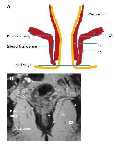
Figure 4 Normal anatomy of lower rectum.
Schematic (A) and coronal plane T2-weighted (B) magnetic resonance imaging presentation. Puborectal sling, the upper portion of the puborectal muscle displaying the uppermost portion of the anal canal (intermittent line). Anal verge is the lowermost portion of the anal canal (line). LA: Levator ani muscle; IS: Internal sphincter; ES: External sphincter.
- Citation: Engin G, Sharifov R. Magnetic resonance imaging for diagnosis and neoadjuvant treatment evaluation in locally advanced rectal cancer: A pictorial review. World J Clin Oncol 2017; 8(3): 214-229
- URL: https://www.wjgnet.com/2218-4333/full/v8/i3/214.htm
- DOI: https://dx.doi.org/10.5306/wjco.v8.i3.214


