Copyright
©The Author(s) 2017.
World J Clin Oncol. Jun 10, 2017; 8(3): 214-229
Published online Jun 10, 2017. doi: 10.5306/wjco.v8.i3.214
Published online Jun 10, 2017. doi: 10.5306/wjco.v8.i3.214
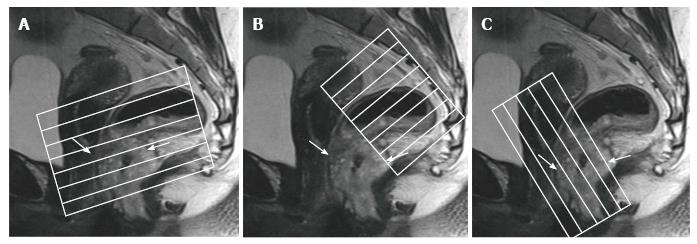
Figure 1 Magnetic resonance imaging planes.
T2-weighted sagittal images are used to determine the longitudinal tumor axis in order to angle the axial and coronal planes. A: Oblique axial plane is obtained perpendicular to the rectal wall at the level of the rectal mass; B: Oblique axial plane is angled perpendicular to the pelvic floor, used to cover lymph node drainage territory; C: Coronal plane is angled parallel to the anal canal for imaging of low rectal tumors. Rectal tumor is indicated by arrows.
- Citation: Engin G, Sharifov R. Magnetic resonance imaging for diagnosis and neoadjuvant treatment evaluation in locally advanced rectal cancer: A pictorial review. World J Clin Oncol 2017; 8(3): 214-229
- URL: https://www.wjgnet.com/2218-4333/full/v8/i3/214.htm
- DOI: https://dx.doi.org/10.5306/wjco.v8.i3.214


