Copyright
©The Author(s) 2017.
World J Clin Oncol. Feb 10, 2017; 8(1): 86-90
Published online Feb 10, 2017. doi: 10.5306/wjco.v8.i1.86
Published online Feb 10, 2017. doi: 10.5306/wjco.v8.i1.86
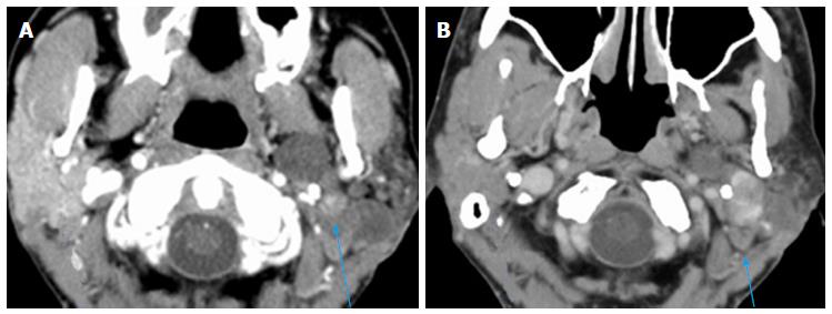
Figure 1 Axial computed tomography of the neck with contrast demonstrates oval shaped enhancing lesion of the left parotid gland deep to the left ramus of the mandible, centered at the left stylomandibular tunnel.
A: The lesion measured 9 mm × 7 mm × 8 mm in 2007; B: The lesion measured 3.1 cm × 2.8 cm × 4.5 cm in 2015.
- Citation: Machado RA, Moubayed SP, Khorsandi A, Hernandez-Prera JC, Urken ML. Intermittent facial spasms as the presenting sign of a recurrent pleomorphic adenoma. World J Clin Oncol 2017; 8(1): 86-90
- URL: https://www.wjgnet.com/2218-4333/full/v8/i1/86.htm
- DOI: https://dx.doi.org/10.5306/wjco.v8.i1.86


