Copyright
©The Author(s) 2016.
World J Clin Oncol. Oct 10, 2016; 7(5): 414-419
Published online Oct 10, 2016. doi: 10.5306/wjco.v7.i5.414
Published online Oct 10, 2016. doi: 10.5306/wjco.v7.i5.414
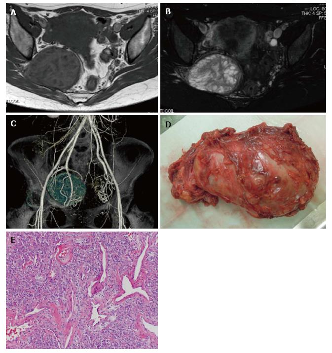
Figure 1 Case 1: An sciatic notch dumbbell tumor in a 41-year-old female.
A and B: Axial MRI revealed an SNDT with a 7.7-cm diameter. The tumor showed a mixed intensity signal on T1- (A) and T2-weighted (B) images; C: 3D-CT angiography clearly demonstrated the relationship between the tumor and major vessels; D: Macroscopic appearance of the resected tumor showing a gray-white, dumbbell-shaped mass with surrounding soft tissue; E: Postoperative pathology confirmed the diagnosis of solitary fibrous tumor. The specimen showed cellular proliferation of mildly atypical spindle or oval cells arranged in short fascicles that were associated with dilated sclerotic blood vessels displaying a hemangiopericytoma-like appearance. Hematoxylin and eosin, original magnification 100 ×. MRI: Magnetic resonance imaging; SNDT: Sciatic notch dumbbell tumor; CT: Computed tomography.
- Citation: Matsumoto Y, Matsunobu T, Harimaya K, Kawaguchi K, Hayashida M, Okada S, Doi T, Iwamoto Y. Bone and soft tissue tumors presenting as sciatic notch dumbbell masses: A critical differential diagnosis of sciatica. World J Clin Oncol 2016; 7(5): 414-419
- URL: https://www.wjgnet.com/2218-4333/full/v7/i5/414.htm
- DOI: https://dx.doi.org/10.5306/wjco.v7.i5.414


