Copyright
©The Author(s) 2016.
World J Clin Oncol. Oct 10, 2016; 7(5): 380-386
Published online Oct 10, 2016. doi: 10.5306/wjco.v7.i5.380
Published online Oct 10, 2016. doi: 10.5306/wjco.v7.i5.380
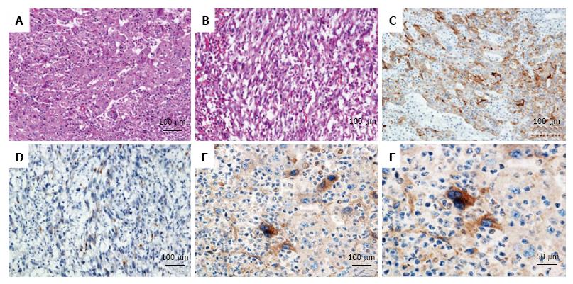
Figure 3 Histopathologic findings.
Microscopic findings showed atypical poorly differentiated cells with a sheet structure (A); HCC tumor was also composed of sarcomatous spindle-shaped cells (B); in both samples, a drastic infiltration of the neutrophils was found (H and E, × 20). Immunohistochemical findings showed CAM5.2 positive in the moderately to poorly differentiated HCC lesion (C) and negative in the spindle-shaped cell lesion (D) (CAM5.2, × 20). Immunohistochemical examination showed that G-CSF was positive in the moderately to poorly differentiated HCC lesion (E, F) (G-CSF, × 20 and × 40). G-CSF: Granulocyte colony-stimulating factor; HCC: Hepatocellular carcinoma cell.
- Citation: Nagata H, Komatsu S, Takaki W, Okayama T, Sawabe Y, Ishii M, Kishimoto M, Otsuji E, Konosu H. Granulocyte colony-stimulating factor-producing hepatocellular carcinoma with abrupt changes. World J Clin Oncol 2016; 7(5): 380-386
- URL: https://www.wjgnet.com/2218-4333/full/v7/i5/380.htm
- DOI: https://dx.doi.org/10.5306/wjco.v7.i5.380


