Copyright
©The Author(s) 2016.
World J Clin Oncol. Oct 10, 2016; 7(5): 380-386
Published online Oct 10, 2016. doi: 10.5306/wjco.v7.i5.380
Published online Oct 10, 2016. doi: 10.5306/wjco.v7.i5.380
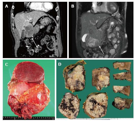
Figure 1 Imaging and macroscopic findings of granulocyte colony-stimulating factor producing hepatocellular carcinoma.
A: CT scan one month before operation showed an irregular liver mass located in segment IV, approximately 60 mm in diameter with peripheral enhancement (white arrow head); B: T2-WI MRI one week before operation showed the rapidly growing liver mass with a 100 mm diameter (white arrow head); C: Macroscopic examination showed a large tumor (100 mm × 100 mm) that protruded through segment IV of the liver to the greater omentum; D: The irregular liver tumor in segment IV showed a central necrosis.
- Citation: Nagata H, Komatsu S, Takaki W, Okayama T, Sawabe Y, Ishii M, Kishimoto M, Otsuji E, Konosu H. Granulocyte colony-stimulating factor-producing hepatocellular carcinoma with abrupt changes. World J Clin Oncol 2016; 7(5): 380-386
- URL: https://www.wjgnet.com/2218-4333/full/v7/i5/380.htm
- DOI: https://dx.doi.org/10.5306/wjco.v7.i5.380


