Copyright
©The Author(s) 2016.
World J Clin Oncol. Jun 10, 2016; 7(3): 308-320
Published online Jun 10, 2016. doi: 10.5306/wjco.v7.i3.308
Published online Jun 10, 2016. doi: 10.5306/wjco.v7.i3.308
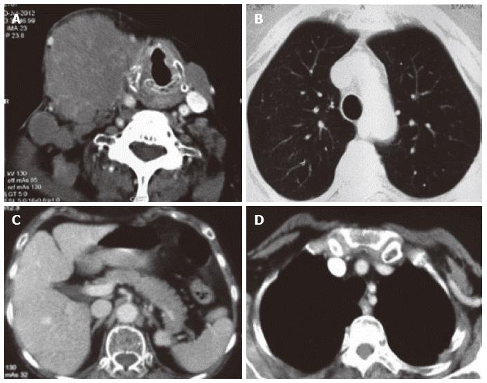
Figure 4 Thoracic imaging at baseline and after treatment of case 2.
A: Contrast enhanced computed tomography of the neck revealing a conglomerate lymph node mass of size 7 cm × 4 cm involving the right submandibular region. The mass is pushing the larynx towards the left side; B: Contrast enhanced computed tomography of the thorax (lung window) with no evidence of primary in the lung; C: Contrast enhanced computed tomography of the abdomen with normal abdominal organs and no evidence of any primary in the abdomen; D: Mediastinal window of CECT thorax revealing no enlarged lymph node stations in the mediastinum. CECT: Contrast enhanced computed tomography.
- Citation: Sehgal IS, Kaur H, Dhooria S, Bal A, Gupta N, Behera D, Singh N. Extrapulmonary small cell carcinoma of lymph node: Pooled analysis of all reported cases. World J Clin Oncol 2016; 7(3): 308-320
- URL: https://www.wjgnet.com/2218-4333/full/v7/i3/308.htm
- DOI: https://dx.doi.org/10.5306/wjco.v7.i3.308


