Copyright
©The Author(s) 2016.
World J Clin Oncol. Jun 10, 2016; 7(3): 308-320
Published online Jun 10, 2016. doi: 10.5306/wjco.v7.i3.308
Published online Jun 10, 2016. doi: 10.5306/wjco.v7.i3.308
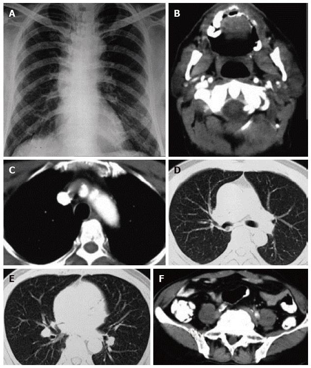
Figure 2 Thoracic imaging at baseline and after treatment of illustrative case 1.
A: Chest radiograph revealing hyperinflated lung fields with no evidence of any parenchymal abnormality; B: Contrast enhanced computed tomography (CECT) of the neck revealing enlarged right sided cervical group of lymph nodes; C: Mediastinal window of CECT of the thorax with no evidence of mediastinal lymph node enlargement; D and E: Lung window of CECT thorax with no evidence of primary in the lung; F: CECT of the abdomen with no evidence of any abnormality in the abdomen.
- Citation: Sehgal IS, Kaur H, Dhooria S, Bal A, Gupta N, Behera D, Singh N. Extrapulmonary small cell carcinoma of lymph node: Pooled analysis of all reported cases. World J Clin Oncol 2016; 7(3): 308-320
- URL: https://www.wjgnet.com/2218-4333/full/v7/i3/308.htm
- DOI: https://dx.doi.org/10.5306/wjco.v7.i3.308


