Copyright
©The Author(s) 2016.
World J Clin Oncol. Jun 10, 2016; 7(3): 293-301
Published online Jun 10, 2016. doi: 10.5306/wjco.v7.i3.293
Published online Jun 10, 2016. doi: 10.5306/wjco.v7.i3.293
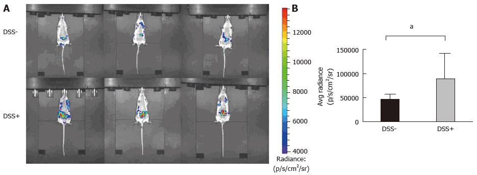
Figure 3 Monitoring of intra-digestive bioluminescent signals using the inflammation probe in the CEABAC10 mouse model.
One mouse group was treated with 1% dextran sodium sulfate (DSS-treated mice group) in drinking water for 6 d (n = 3), while the other group received only drinking water (n = 3; DSS-). Mice were subjected to one cycle of DSS and then imaging with the inflammation probe before sacrifice. Mice were intravenously injected with the inflammation probe, and the BLI signal was detected immediately after injection. DSS-treated mouse group acquisition (A) showed severe intra-digestive inflammation was correlated with a significant increase in the BLI signal on graphs (B) compared to the DSS group (aP = 0.03). BLI: Bioluminescent; DSS: Dextran sodium sulfate.
- Citation: Veziant J, Gagnière J, Jouberton E, Bonnin V, Sauvanet P, Pezet D, Barnich N, Miot-Noirault E, Bonnet M. Association of colorectal cancer with pathogenic Escherichia coli: Focus on mechanisms using optical imaging. World J Clin Oncol 2016; 7(3): 293-301
- URL: https://www.wjgnet.com/2218-4333/full/v7/i3/293.htm
- DOI: https://dx.doi.org/10.5306/wjco.v7.i3.293


