Copyright
©The Author(s) 2016.
World J Clin Oncol. Jun 10, 2016; 7(3): 293-301
Published online Jun 10, 2016. doi: 10.5306/wjco.v7.i3.293
Published online Jun 10, 2016. doi: 10.5306/wjco.v7.i3.293
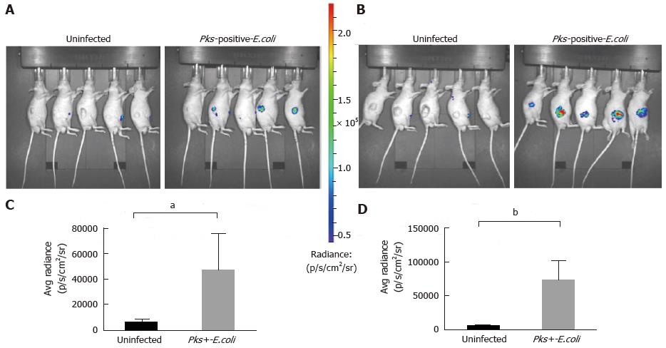
Figure 2 Significant increase of inflammation in HCT116 tumors infected with Escherichia coli strains measured by optical imaging using the inflammation probe.
In vivo BLI imaging was performed at day 20 (A) and day 34 (B) post-HCT116 xenograft in nude mice (n = 5 animals with uninfected xenograft; n = 5 animals in pks-positive E. coli-infected xenograft). BLI images comparing uninfected to pks-positive E. coli-infected xenografts are shown on day 20 (A) and day 34 (B) post-xenograft. Intensity of emission is represented as the pseudocolor image. A sharp increase in the BLI signal was seen in the pks-positive E. coli-infected xenograft group at each time point. The BLI signal detected 10 min after i.p. probe injection was quantified from the ROI drawn manually. Photon flux was expressed as the average radiance in p/s/cm2/sr. Graphs reveal a statistically significant increase in the BLI signal in the pks-positive E. coli-infected tumors: day 20, aP = 0.0132 (C) and day 34, bP = 0.0006 (D). BLI: Bioluminescent; E. coli: Escherichia coli; i.p.: Intraperitoneal.
- Citation: Veziant J, Gagnière J, Jouberton E, Bonnin V, Sauvanet P, Pezet D, Barnich N, Miot-Noirault E, Bonnet M. Association of colorectal cancer with pathogenic Escherichia coli: Focus on mechanisms using optical imaging. World J Clin Oncol 2016; 7(3): 293-301
- URL: https://www.wjgnet.com/2218-4333/full/v7/i3/293.htm
- DOI: https://dx.doi.org/10.5306/wjco.v7.i3.293


