Copyright
©The Author(s) 2015.
World J Clin Oncol. Aug 10, 2015; 6(4): 57-63
Published online Aug 10, 2015. doi: 10.5306/wjco.v6.i4.57
Published online Aug 10, 2015. doi: 10.5306/wjco.v6.i4.57
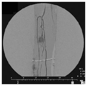
Figure 2 Angiogram of melanoma.
In a patient with extremity melanoma AJCC stage IIIC, angiogram shows in the middle third of the leg rounded nodule with a homogeneous, hypervascular stain, nourished by muscular branches of the peroneal artery. Additional deep-seated lesions, which were not palpable clinically, were detected by angiography distally in the leg, nourished by branches of the anterior tibial artery.
- Citation: Cecchini S, Sarti D, Ricci S, Vergini LD, Sallei M, Serresi S, Ricotti G, Mulazzani L, Lattanzio F, Fiorentini G. Isolated limb infusion chemotherapy with or without hemofiltration for recurrent limb melanoma. World J Clin Oncol 2015; 6(4): 57-63
- URL: https://www.wjgnet.com/2218-4333/full/v6/i4/57.htm
- DOI: https://dx.doi.org/10.5306/wjco.v6.i4.57


