Copyright
©2014 Baishideng Publishing Group Co.
World J Clin Oncol. May 10, 2014; 5(2): 61-70
Published online May 10, 2014. doi: 10.5306/wjco.v5.i2.61
Published online May 10, 2014. doi: 10.5306/wjco.v5.i2.61
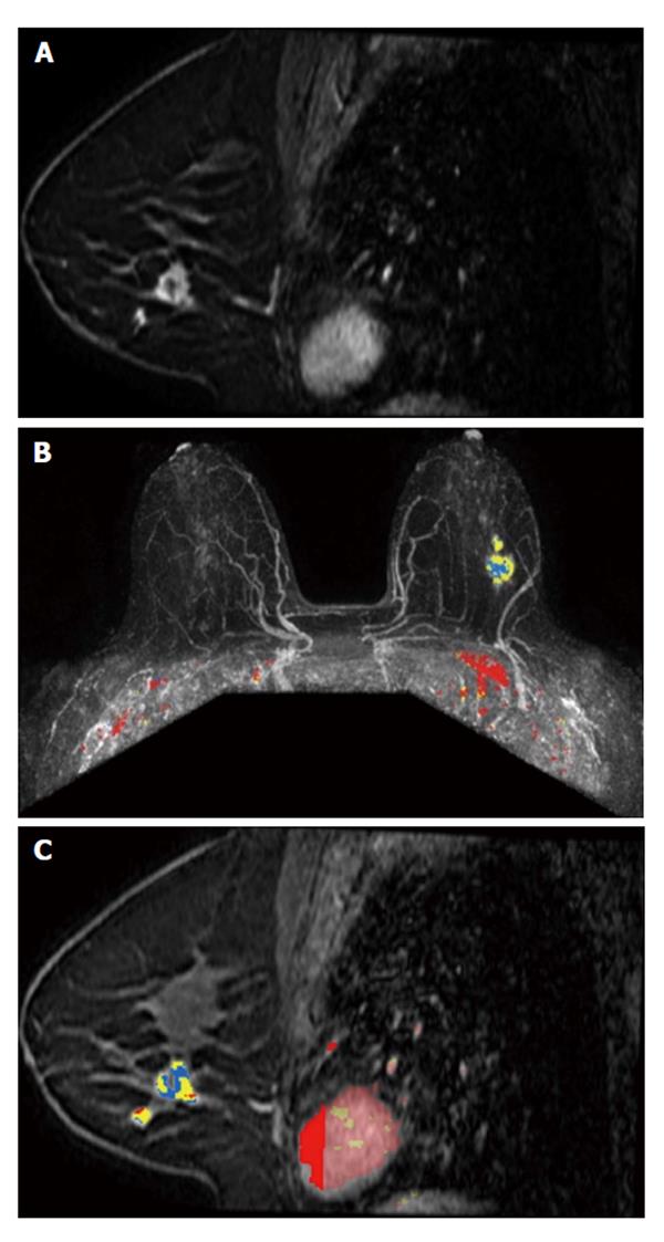
Figure 2 A 48-year-old woman, with positive family history for breast cancer, presented with a palpable lump on the left breast, finally diagnosed as invasive ductolobular carcinoma.
A: Sagittal contrast-enhanced fat-suppressed T1-weighted gradient echo images obtained at 3 T shows a spiculated mass, with rim enhancement and small satellite lesion (multifocal disease); B and C: Color parametric enhancement map in axial postcontrast maximum intensity projection and sagittal projection indicates predominantly a plateau enhancement behavior, with some areas of washout.
- Citation: Menezes GL, Knuttel FM, Stehouwer BL, Pijnappel RM, van den Bosch MA. Magnetic resonance imaging in breast cancer: A literature review and future perspectives. World J Clin Oncol 2014; 5(2): 61-70
- URL: https://www.wjgnet.com/2218-4333/full/v5/i2/61.htm
- DOI: https://dx.doi.org/10.5306/wjco.v5.i2.61


