Copyright
©2014 Baishideng Publishing Group Co.
World J Clin Oncol. Feb 10, 2014; 5(1): 28-32
Published online Feb 10, 2014. doi: 10.5306/wjco.v5.i1.28
Published online Feb 10, 2014. doi: 10.5306/wjco.v5.i1.28
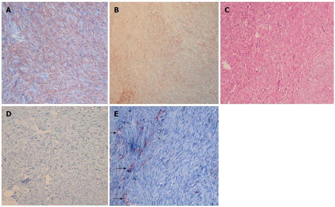
Figure 4 Diagnosis of metastatic retroperitoneal leiomyosarcoma.
A: Strong positive SMA immunohistochemical staining in clitoral tumor (× 100); B: Immunohistochemically, spindle-shape tumour cells revealed staining for desmin in the clitoral tumor (× 100); C: Spindle shaped tumour cells with eosinophilic cytoplasm and nuclear polymorphism in retroperitoneal leimyosarcoma (× 100 magnification, Haemaetoxylin-Eosin staining); D: The retroperitoneal tumour cells were negative for desmin in retroperitoneal leiomyosarcoma (× 100); E: Immunohistochemistry reveals CD34 negative tumour cells and CD34 positive vascular endothelial cells (arrows) in retroperitoneal leiomyosarcoma (× 200).
- Citation: Cokmert S, Demir L, Akyol M, Bayoglu IV, Can A, Unek IT, Bolat FA. Clitoris metastasis from a retroperitoneal leiomyosarcoma: A case report. World J Clin Oncol 2014; 5(1): 28-32
- URL: https://www.wjgnet.com/2218-4333/full/v5/i1/28.htm
- DOI: https://dx.doi.org/10.5306/wjco.v5.i1.28


