Copyright
©2013 Baishideng Publishing Group Co.
World J Clin Oncol. Nov 10, 2013; 4(4): 91-101
Published online Nov 10, 2013. doi: 10.5306/wjco.v4.i4.91
Published online Nov 10, 2013. doi: 10.5306/wjco.v4.i4.91
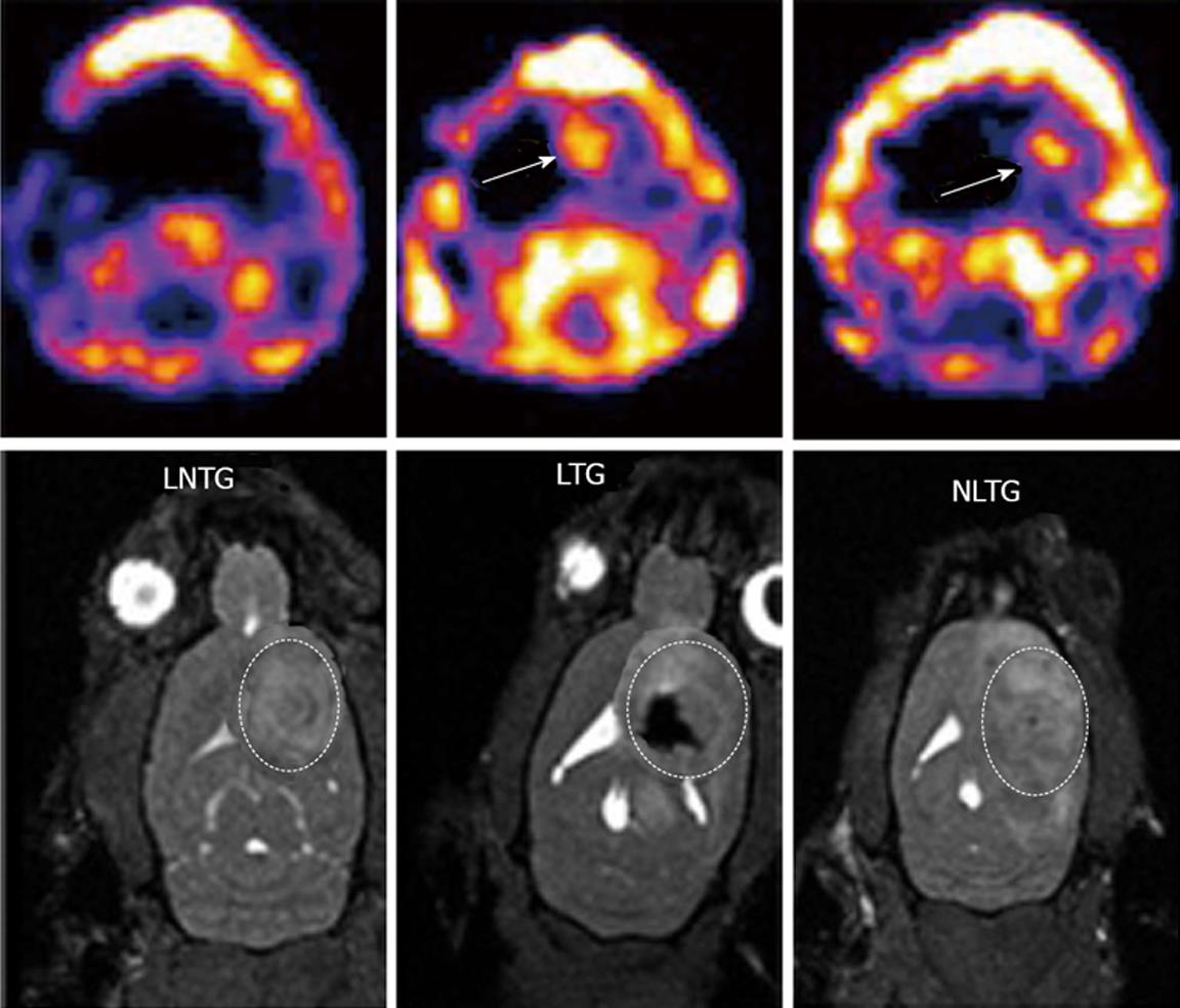
Figure 2 Magnetic resonance imaging and single photon emission computed tomography images for tracking of intratumor injected endothelial progenitor cells and transgene expression.
Expression of transgene (hNIS) was detected by single photon emission computed tomography (SPECT) and magnetic resonance imaging (MRI) was used to detect the cell migration in the tumor. Upper panels (SPECT images): Left: Animals received non transgenic, non-labeled endothelial progenitor cells (EPCs) (control); Middle: Animals received transgenic EPCs labeled with iron; Right: Animals received non labeled transgenic EPCs. Corresponding MRI images are shown in the lower panels. Animals received labeled transgenic EPC or non-labeled transgenic EPC showed higher activities of Tc-99m in the tumors compared to control animals that received non-transgenic EPCs. MRI images clearly indicate the presence of iron labeled transgenic EPCs (lower panel, middle).
- Citation: Varma NRS, Barton KN, Janic B, Shankar A, Iskander A, Ali MM, Arbab AS. Monitoring adenoviral based gene delivery in rat glioma by molecular imaging. World J Clin Oncol 2013; 4(4): 91-101
- URL: https://www.wjgnet.com/2218-4333/full/v4/i4/91.htm
- DOI: https://dx.doi.org/10.5306/wjco.v4.i4.91


