Copyright
©2013 Baishideng Publishing Group Co.
World J Clin Oncol. Nov 10, 2013; 4(4): 102-105
Published online Nov 10, 2013. doi: 10.5306/wjco.v4.i4.102
Published online Nov 10, 2013. doi: 10.5306/wjco.v4.i4.102
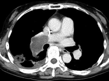
Figure 1 Axial enhanced computed tomography.
At the level of the bronchus intermedius, showing a filling defect that occupies the entire lumina of the right and interlobar pulmonary arteries.
- Citation: Fukai R, Rokkaku K, Irie Y, Imazeki T, Katada Y, Watanabe H, Ueda Y, Miyamoto H, Chida M. Pulmonary artery sarcoma successfully treated by right pneumonectomy after definitive diagnosis. World J Clin Oncol 2013; 4(4): 102-105
- URL: https://www.wjgnet.com/2218-4333/full/v4/i4/102.htm
- DOI: https://dx.doi.org/10.5306/wjco.v4.i4.102


