Copyright
©2013 Baishideng Publishing Group Co.
World J Clin Oncol. Aug 10, 2013; 4(3): 75-81
Published online Aug 10, 2013. doi: 10.5306/wjco.v4.i3.75
Published online Aug 10, 2013. doi: 10.5306/wjco.v4.i3.75
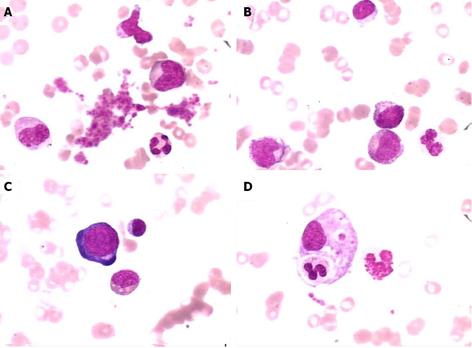
Figure 3 Representative images of bone marrow examination.
The images showed anemia with marked red cell aplasia and the remarkable proliferation of granulocytes and megakaryocytes (Wright and Giemsa stain, × 1000). A: platelet; B: phagocyte; C: pronormoblast; D: megakaryocyte.
- Citation: Tao J, Zheng FP, Tian H, Lin Y, Li JZ, Chen XL, Chen JN, Shao CK, Wu B. Angioimmunoblastic T-cell lymphoma-associated pure red cell aplasia with abdominal pain. World J Clin Oncol 2013; 4(3): 75-81
- URL: https://www.wjgnet.com/2218-4333/full/v4/i3/75.htm
- DOI: https://dx.doi.org/10.5306/wjco.v4.i3.75


