Copyright
©2013 Baishideng.
World J Clin Oncol. Feb 10, 2013; 4(1): 25-28
Published online Feb 10, 2013. doi: 10.5306/wjco.v4.i1.25
Published online Feb 10, 2013. doi: 10.5306/wjco.v4.i1.25
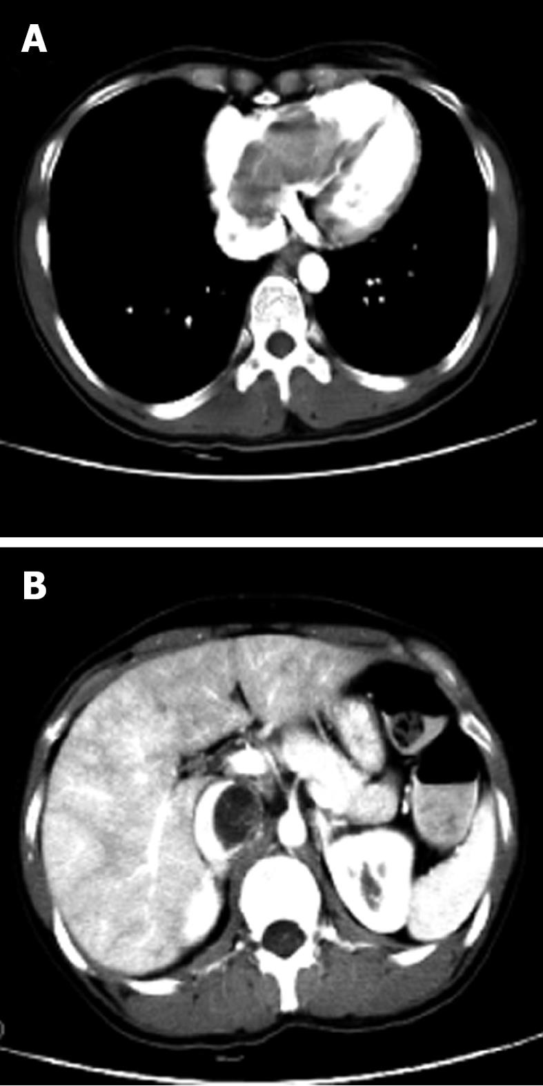
Figure 3 Preoperative chest and abdominal computed tomography.
A: Axial image demonstrates a lobulated mass within the right atrium and ventricle; B: Axial image shows a tumor in the inferior vena cava and resulting lumen stenosis.
- Citation: Xu ZF, Yong F, Chen YY, Pan AZ. Uterine intravenous leiomyomatosis with cardiac extension: Imaging characteristics and literature review. World J Clin Oncol 2013; 4(1): 25-28
- URL: https://www.wjgnet.com/2218-4333/full/v4/i1/25.htm
- DOI: https://dx.doi.org/10.5306/wjco.v4.i1.25


