Copyright
©2012 Baishideng Publishing Group Co.
World J Clin Oncol. Jun 10, 2012; 3(6): 92-97
Published online Jun 10, 2012. doi: 10.5306/wjco.v3.i6.92
Published online Jun 10, 2012. doi: 10.5306/wjco.v3.i6.92
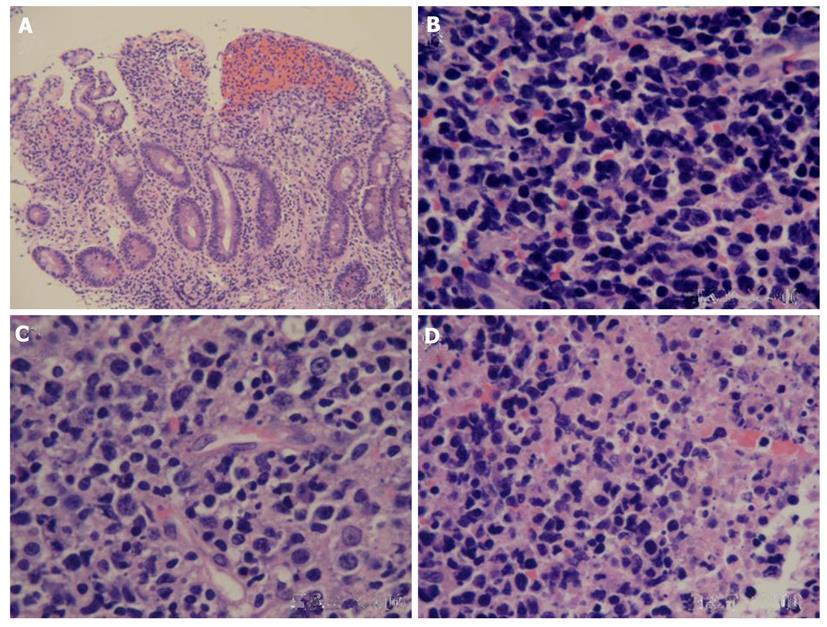
Figure 3 Photomicrograph of biopsy specimens.
A: A large numbers of atypical cells infiltrated the mucosa and mucosal glands became widely spaced or lost [hematoxylin and eosin (HE) stain , × 100]; B: The tumor was composed of medium-sized cells (HE stain, × 400); C: The large cells admixed with small cells (HE stain, × 400); D: Coagulative necrosis and admixed apoptotic bodies were observed in the specimens (HE stain, × 400).
- Citation: Li JZ, Tao J, Ruan DY, Yang YD, Zhan YS, Wang X, Chen Y, Kuang SC, Shao CK, Wu B. Primary duodenal NK/T-cell lymphoma with massive bleeding: A case report. World J Clin Oncol 2012; 3(6): 92-97
- URL: https://www.wjgnet.com/2218-4333/full/v3/i6/92.htm
- DOI: https://dx.doi.org/10.5306/wjco.v3.i6.92


