Copyright
©2012 Baishideng Publishing Group Co.
World J Clin Oncol. Dec 10, 2012; 3(12): 155-158
Published online Dec 10, 2012. doi: 10.5306/wjco.v3.i12.155
Published online Dec 10, 2012. doi: 10.5306/wjco.v3.i12.155
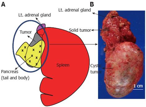
Figure 3 The schema shows the macroscopic appearance of the resected specimen.
A: The cystic tumor (circled in blue) was separated from the pancreas by a fibrous tumor membrane; B: The tumor measured 10 cm × 4 cm in diameter and was filled with homogenous, grayish, creamy fluid. This tumor also had a solid component measuring 3 cm × 2.5 cm in diameter adjacent to the left adrenal gland.
- Citation: Hayama S, Ohmi M, Yonemori A, Yamabuki T, Inomata H, Nihei K, Hirano S. Ganglioneuroblastoma arising within a retroperitoneal mature cystic teratoma. World J Clin Oncol 2012; 3(12): 155-158
- URL: https://www.wjgnet.com/2218-4333/full/v3/i12/155.htm
- DOI: https://dx.doi.org/10.5306/wjco.v3.i12.155


