Copyright
©2012 Baishideng Publishing Group Co.
World J Clin Oncol. Jan 10, 2012; 3(1): 7-11
Published online Jan 10, 2012. doi: 10.5306/wjco.v3.i1.7
Published online Jan 10, 2012. doi: 10.5306/wjco.v3.i1.7
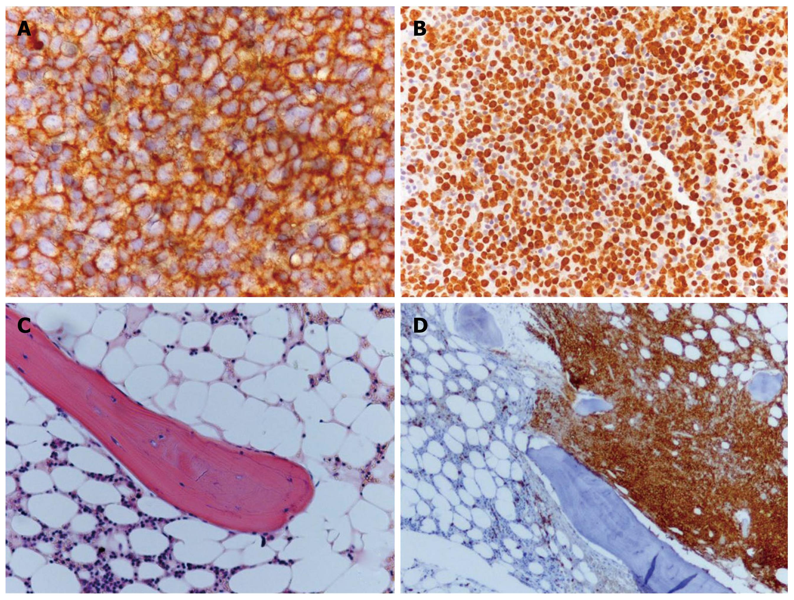
Figure 2 Histological sections of axillary lymph node (A and B) and bone marrow (C and D).
Lymph node: A: Immunohistochemical stain for CD20; B: Immunohistochemical stain for Ki67; Bone marrow: C: Hematoxylin-eosin staining; D: Immunohistochemical stain for CD20.
- Citation: Pellicciotti F, Giusti A, Gelli MC, Foderaro S, Ferrari A, Pioli G. Challenges in the differential diagnosis of hypercalcemia: A case of hypercalcemia with normal PTH level. World J Clin Oncol 2012; 3(1): 7-11
- URL: https://www.wjgnet.com/2218-4333/full/v3/i1/7.htm
- DOI: https://dx.doi.org/10.5306/wjco.v3.i1.7


