Copyright
©2011 Baishideng Publishing Group Co.
World J Clin Oncol. May 10, 2011; 2(5): 217-224
Published online May 10, 2011. doi: 10.5306/wjco.v2.i5.217
Published online May 10, 2011. doi: 10.5306/wjco.v2.i5.217
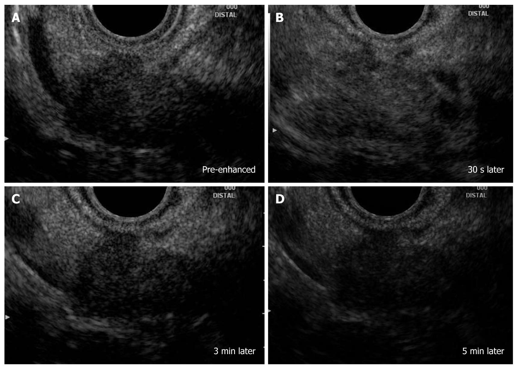
Figure 6 Pancreatic ductal adenocarcinoma.
A: There was a hypoechoic mass at the pancreatic body with an irregular margin (pre-enhanced); B: The image indicated an increase in echo-intensity close to that of the surrounding normal parenchyma (30 s later after injection of Sonazoid); C, D: The center and the rightmost images were obtained 3 min and 5 min after injection. The tumor became hypoechoic (i.e. hypovascular) compared with surrounding tissues.
- Citation: Hirooka Y, Itoh A, Kawashima H, Ohno E, Ishikawa T, Itoh Y, Nakamura Y, Hiramatsu T, Nakamura M, Miyahara R, Ohmiya N, Ishigami M, Katano Y, Goto H. Clinical oncology for pancreatic and biliary cancers: Advances and current limitations. World J Clin Oncol 2011; 2(5): 217-224
- URL: https://www.wjgnet.com/2218-4333/full/v2/i5/217.htm
- DOI: https://dx.doi.org/10.5306/wjco.v2.i5.217


