Copyright
©2011 Baishideng Publishing Group Co.
World J Clin Oncol. May 10, 2011; 2(5): 217-224
Published online May 10, 2011. doi: 10.5306/wjco.v2.i5.217
Published online May 10, 2011. doi: 10.5306/wjco.v2.i5.217
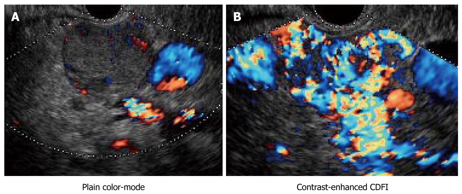
Figure 5 Endocrine tumor of the pancreas.
A: Plain color (non-enhanced color) Doppler endoscopic ultrasonography (EUS) only showed few color signals inside the clearly delineated iso-echoic tumor; B: In contrast, there were abundant color signals inside the tumor, indicating it was hypervascular. B-mode image combined with contrast-enhanced EUS images provided an important clue for the diagnosis of pancreatic endocrine tumors. Levovist® was used in this case.
- Citation: Hirooka Y, Itoh A, Kawashima H, Ohno E, Ishikawa T, Itoh Y, Nakamura Y, Hiramatsu T, Nakamura M, Miyahara R, Ohmiya N, Ishigami M, Katano Y, Goto H. Clinical oncology for pancreatic and biliary cancers: Advances and current limitations. World J Clin Oncol 2011; 2(5): 217-224
- URL: https://www.wjgnet.com/2218-4333/full/v2/i5/217.htm
- DOI: https://dx.doi.org/10.5306/wjco.v2.i5.217


