Copyright
©2011 Baishideng Publishing Group Co.
World J Clin Oncol. Apr 10, 2011; 2(4): 187-194
Published online Apr 10, 2011. doi: 10.5306/wjco.v2.i4.187
Published online Apr 10, 2011. doi: 10.5306/wjco.v2.i4.187
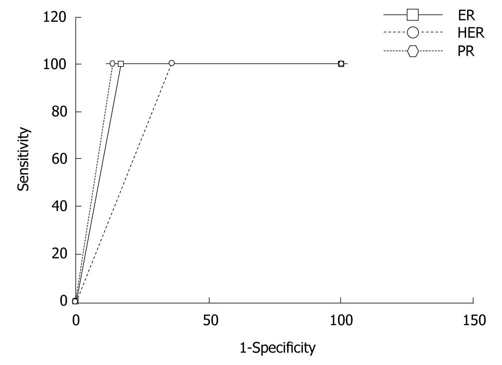
Figure 2 ROC curves for the automated evaluation of estrogen receptor, progesterone receptor and human epidermal growth factor receptor-2 expression images using TissueQuant.
For all three expressions the sensitivity of 100% is maintained, the specificity for PR expression is best at, 86%, for ER the specificity is 82.3% and for HER-2/neu expression the specificity is least at, 64.3%. ER: Estrogen receptor; PR: Progesterone receptor; HER-2/neu: Human epidermal growth factor receptor-2.
- Citation: Prasad K, Tiwari A, Ilanthodi S, Prabhu G, Pai M. Automation of immunohistochemical evaluation in breast cancer using image analysis. World J Clin Oncol 2011; 2(4): 187-194
- URL: https://www.wjgnet.com/2218-4333/full/v2/i4/187.htm
- DOI: https://dx.doi.org/10.5306/wjco.v2.i4.187


