Copyright
©2011 Baishideng Publishing Group Co.
World J Clin Oncol. Jan 10, 2011; 2(1): 8-27
Published online Jan 10, 2011. doi: 10.5306/wjco.v2.i1.8
Published online Jan 10, 2011. doi: 10.5306/wjco.v2.i1.8
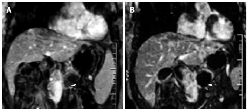
Figure 6 Dynamic contrast-enhanced gradient-echo T1-weighted magnetic resonance images (180/6.
0, 90° flip angle, 128 × 256 matrix, 10-mm-thick sections, 2-mm intersection gap, one signal acquired, and 18-s acquisition time) obtained with breath holding in 48-year-old man who underwent high-intensity focused ultrasound ablation for advanced pancreatic cancer. The tumor was 4.5 cm × 4.5 cm in diameter and located in the body of the pancreas. A: Image obtained before high-intensity focused ultrasound shows the blood supply in the pancreatic lesion (arrowhead); B: Image obtained 2 wk after high-intensity focused ultrasound shows no evidence of contrast enhancement in the treated lesion (arrowhead), which is indicative of complete coagulation necrosis in the pancreatic cancer.
- Citation: Zhou YF. High intensity focused ultrasound in clinical tumor ablation. World J Clin Oncol 2011; 2(1): 8-27
- URL: https://www.wjgnet.com/2218-4333/full/v2/i1/8.htm
- DOI: https://dx.doi.org/10.5306/wjco.v2.i1.8


