Copyright
©2011 Baishideng Publishing Group Co.
World J Clin Oncol. Jan 10, 2011; 2(1): 8-27
Published online Jan 10, 2011. doi: 10.5306/wjco.v2.i1.8
Published online Jan 10, 2011. doi: 10.5306/wjco.v2.i1.8
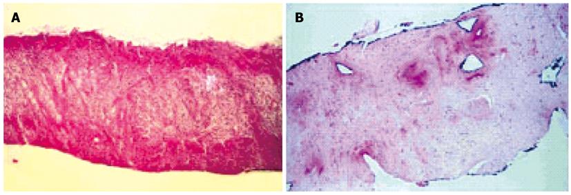
Figure 3 Complete destruction of the glandular tissue due to coagulation necrosis lesion which reaches the capsula and the periprostatic fat 48 h after high intensity focused ultrasound treatment (A) and the necrotic prostatic tissue is replaced by a fibrotic tissue, including the capsula, 3 mo after high intensity focused ultrasound therapy (B).
- Citation: Zhou YF. High intensity focused ultrasound in clinical tumor ablation. World J Clin Oncol 2011; 2(1): 8-27
- URL: https://www.wjgnet.com/2218-4333/full/v2/i1/8.htm
- DOI: https://dx.doi.org/10.5306/wjco.v2.i1.8


