Copyright
©The Author(s) 2025.
World J Clin Oncol. Apr 24, 2025; 16(4): 103564
Published online Apr 24, 2025. doi: 10.5306/wjco.v16.i4.103564
Published online Apr 24, 2025. doi: 10.5306/wjco.v16.i4.103564
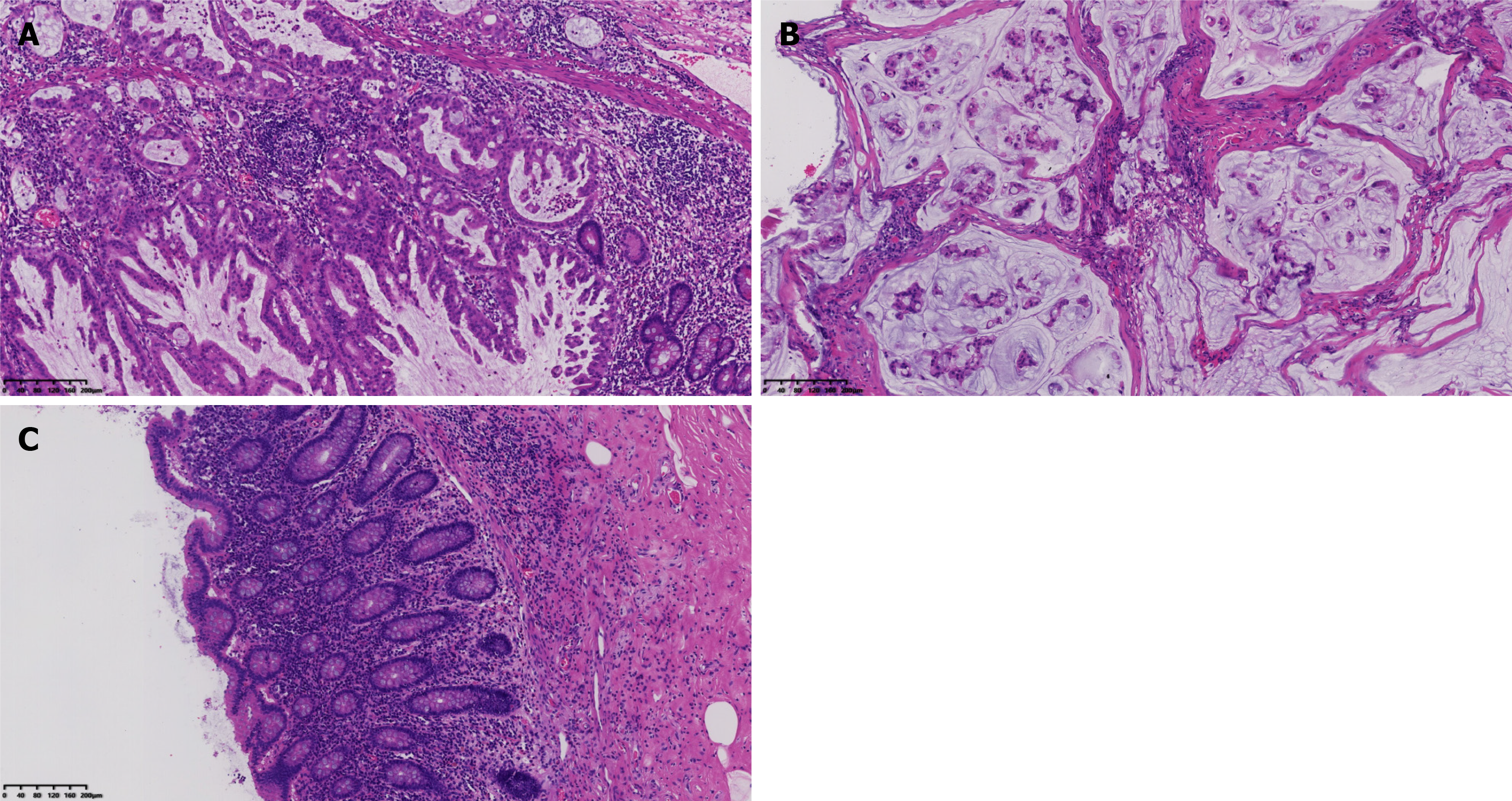
Figure 2 Pathological examinations.
A: Serrated lesions and high-grade dysplasia can be seen in the mucosa of the jejunum (× 100); B: Mucin lakes and severely dysmorphic mucus glands in the peritoneal fibrous tissue (× 100); C Normal appendix structure. Hematoxylin-eosin staining (× 100).
- Citation: Shi GJ, Wang C, Zhang P, Lu YY, Zhou HP, Ma RQ, An LB. Pseudomyxoma peritonei originating from small intestine: A case report and review of literature. World J Clin Oncol 2025; 16(4): 103564
- URL: https://www.wjgnet.com/2218-4333/full/v16/i4/103564.htm
- DOI: https://dx.doi.org/10.5306/wjco.v16.i4.103564


