Copyright
©The Author(s) 2025.
World J Clin Oncol. Apr 24, 2025; 16(4): 102297
Published online Apr 24, 2025. doi: 10.5306/wjco.v16.i4.102297
Published online Apr 24, 2025. doi: 10.5306/wjco.v16.i4.102297
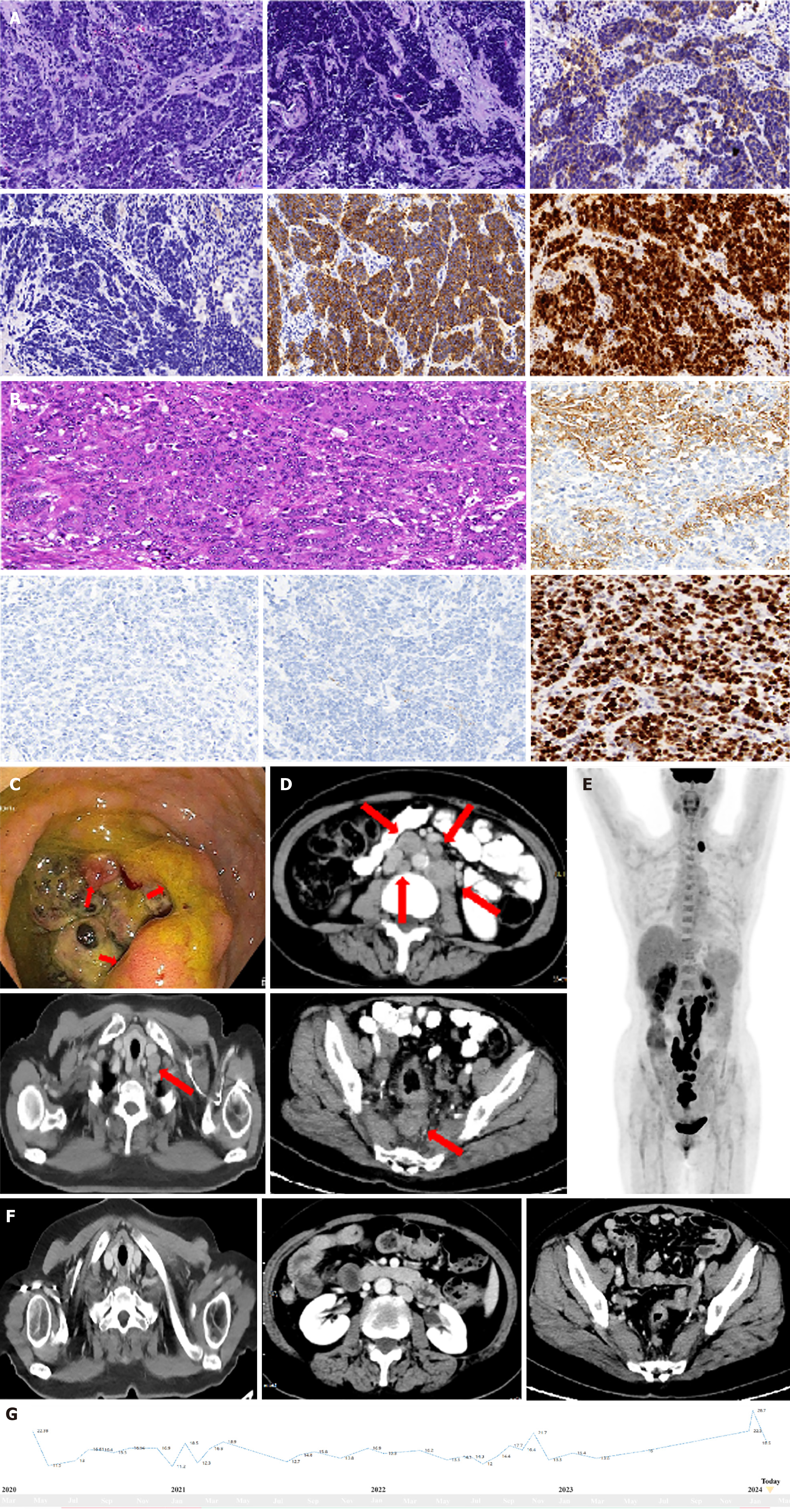
Figure 1 Imaging examination, laboratory examination for cases.
A: HE staining revealed poorly differentiated atypical cells with mixed large and small cells; the scale bar at the bottom right represents 1 cm. Immunohistochemistry of Syn, CgA, CD56 and ki-67; B: Pathological assessment of surgical tissue slides: HE staining revealed poorly differentiated ulcerous mixed neuroendocrine/neuroendocrine carcinoma (MiNEC), with a ratio of 30% poorly differentiated adenocarcinoma to MiNEC; C: Colonoscopy revealed a circumferential rectal neoplasm with an irregular shape; D: Computed tomography (CT) at diagnosis revealed irregular thickening of the rectal wall with uneven enhancement and multiple enlarged nodes in the pelvic cavity, abdomen, and left neck; E: PET/CT at diagnosis revealed hypermetabolic nodes in the pelvic cavity, abdomen, and left neck; F: CTs after 11 cycles of camrelizumab and surufatinib revealed partial remission; the sizes of both the primary rectal lesion and the lymph nodes had decreased (more than 30%); G: Dynamics of NSE during the treatment process.
- Citation: Gao LL, Gao DN, Yuan HT, Chen WQ, Yang J, Peng JQ. Combining anti-PD-1 antibodies with surufatinib for gastrointestinal neuroendocrine carcinoma: Two cases report and review of literature. World J Clin Oncol 2025; 16(4): 102297
- URL: https://www.wjgnet.com/2218-4333/full/v16/i4/102297.htm
- DOI: https://dx.doi.org/10.5306/wjco.v16.i4.102297


