Copyright
©The Author(s) 2024.
World J Clin Oncol. Sep 24, 2024; 15(9): 1207-1214
Published online Sep 24, 2024. doi: 10.5306/wjco.v15.i9.1207
Published online Sep 24, 2024. doi: 10.5306/wjco.v15.i9.1207
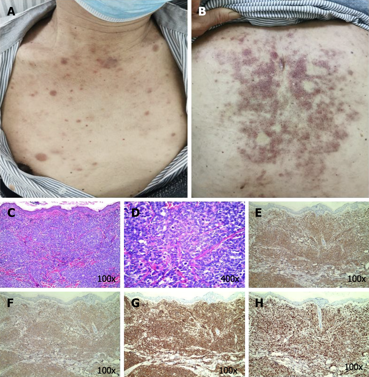
Figure 1 Skin lesions and the images of the hematoxylin-eosin and immunohistochemistry analyses for patient 1.
The image above depicts multiple nodules and bruise-like lesions on the chest and back. A: There is an evident acellular infiltration zone between the tumor cells and the epidermis [hematoxylin-eosin (HE) staining, 100 ×]; B: Microscopically, the dermis is diffused with infiltrated medium-sized blast cell (HE staining, 400 ×); C: CD123 positive; D: CD4 positive; E: CD56 positive; F: Approximately 75% of Ki67 were positive; G: CD56 positive; H: Approximately 75% of Ki67 were positive.
- Citation: Ma YQ, Sun Z, Li YM, Xu H. Blastic plasmacytoid dendritic cell neoplasm: Two case reports. World J Clin Oncol 2024; 15(9): 1207-1214
- URL: https://www.wjgnet.com/2218-4333/full/v15/i9/1207.htm
- DOI: https://dx.doi.org/10.5306/wjco.v15.i9.1207


