Copyright
©The Author(s) 2024.
World J Clin Oncol. Aug 24, 2024; 15(8): 1102-1109
Published online Aug 24, 2024. doi: 10.5306/wjco.v15.i8.1102
Published online Aug 24, 2024. doi: 10.5306/wjco.v15.i8.1102
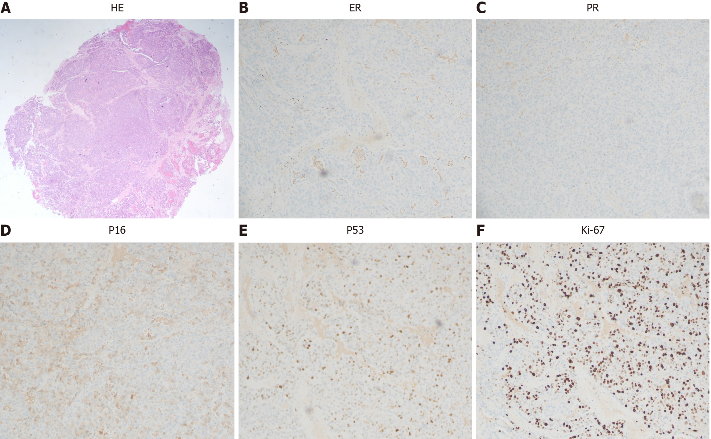
Figure 4 Histological and immunohistochemical examinations.
A: Hematoxylin eosin staining (20 × microscopy); B: Estrogen receptor (5% +, 100 × microscopy); C: Progesterone receptor (−, 100 × microscopy); D: P16 (some weak +, 100 × microscopy); E: P53 (patchy +) (100 × microscopy); F: Ki-67 (70% +, 100 × microscopy).
- Citation: Saijilafu, Gu YJ, Huang AW, Xu CF, Qian LW. Individualized vaginal applicator for stage IIb primary vaginal adenocarcinoma: A case report. World J Clin Oncol 2024; 15(8): 1102-1109
- URL: https://www.wjgnet.com/2218-4333/full/v15/i8/1102.htm
- DOI: https://dx.doi.org/10.5306/wjco.v15.i8.1102


